
Blood vessel abnormalities: orbital varices
Orbital varices represent the most frequent abnormality of the blood vessels in the orbit of the eye. In most cases they affect one half of the face and are located in the supero-nasal quadrant of the eye
What are orbital varices?
The disease is characterised by dilatation of one or more venous vessels (‘caput medusae’).
If localised in the eyelids or under the conjunctiva, the dilatation may be visible. Often the problem may extend to the orbit.
Possible complications include haemorrhage and thrombosis.
What are the causes of orbital varices?
Varicose veins can form due to a weakness in the vessel wall of congenital origin or due to factors such as compression from a tumour, an arterial aneurysm, an arteriovenous malformation, trauma or an infection involving the vein wall.
Another possible cause, present alone or at the same time as the weakness of the vessel wall, can be obstruction of a vein.
What are the symptoms of orbital varices?
The first symptom of an orbital varicose vein is a protrusion of the eyeball that is not pulsating, not associated with blowing and intermittent.
In fact, since the veins of the orbit are valveless, the protrusion is reversible.
It is caused or aggravated by increased venous pressure, associated, for example, with a cough, exertion or forced exhalation with the glottis closed (the so-called Valsava manoeuvre).
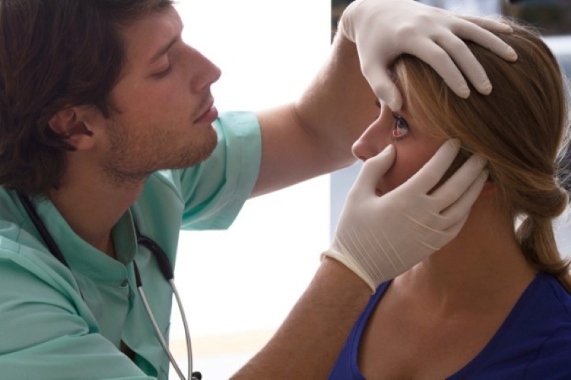
Diagnosis
Diagnosis is clinical and the Valsalva manoeuvre (forced exhalation with the glottis closed) is essential for making the diagnosis.
The examinations that may be prescribed are:
- Visual field examination
- Orthoptic examination
- Nuclear magnetic resonance imaging of the orbits to study the extent of the lesion within them
- Multidisciplinary consultation
Treatments
Treatment is surgical, involves ‘occlusion’ of the varices and is indicated in cases of recurrent thrombosis, pain, severe eyeball protrusion and optic nerve compression.
The operation is complex and often incompletely performed because these lesions are fragile and bleed easily.
Prevention

Unfortunately, there are no preventive measures.
Read Also:
Emergency Live Even More…Live: Download The New Free App Of Your Newspaper For IOS And Android
Dry Eye Syndrome: Symptoms, Causes And Remedies
Red Eyes: What Can Be The Causes Of Conjunctival Hyperemia?
Autoimmune Diseases: The Sand In The Eyes Of Sjögren’s Syndrome
Corneal Abrasions And Foreign Bodies In The Eye: What To Do? Diagnosis And Treatment
Covid, A ‘Mask’ For The Eyes Thanks To Ozone Gel: An Ophthalmic Gel Under Study
Dry Eyes In Winter: What Causes Dry Eye In This Season?
What Is Aberrometry? Discovering The Aberrations Of The Eye
Raising The Bar For Pediatric Trauma Care: Analysis And Solutions In The US
What Is Ocular Pressure And How Is It Measured?
Opening The Eyes Of The World, CUAMM’s “ForeSeeing Inclusion” Project To Combat Blindness In Uganda
What Is Ocular Myasthenia Gravis And How Is It Treated?
Retinal Detachment: When To Worry About Myodesopias, The ‘Flying Flies’
Retinal Thrombosis: Symptoms, Diagnosis And Treatment Of Retinal Vessel Occlusion
My Eye Dances: Learning About Nystagmus