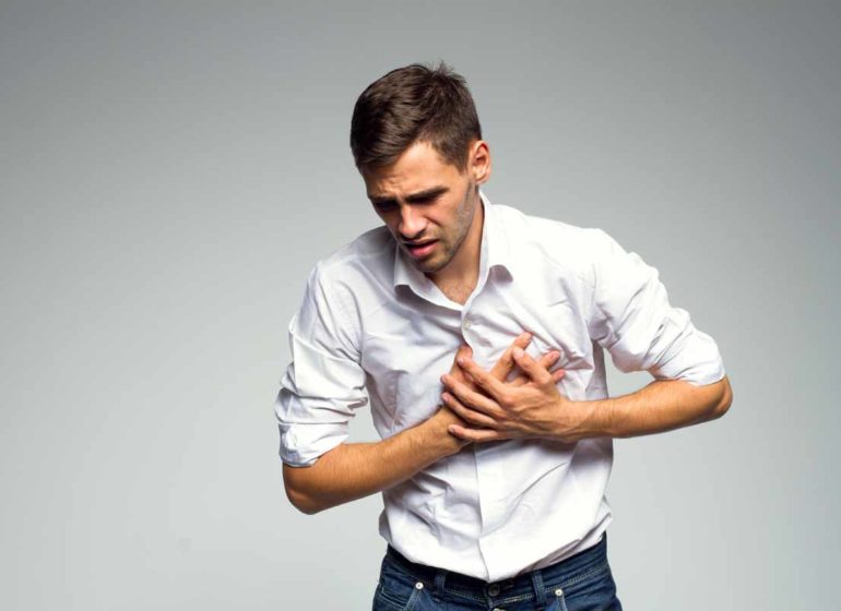
A feeling of tightness in the chest? Could be angina pectoris
Angina is chest pain or a feeling of pressure that is felt when the heart is not getting the right amount of oxygen
If you have angina, you feel a sensation of discomfort under your sternum. Angina is felt when exerting oneself and subsides at rest.
In order to diagnose angina, in addition to the symptoms, an electrocardiogram and other diagnostic tests are performed.
Treatment ranges from the administration of beta blockers, calcium channel blockers, to percutaneous coronary intervention or coronary artery bypass grafting.
Causes of angina
The heart needs a constant supply of blood and oxygen; ensuring this are the coronary arteries that branch off the aorta as it exits the heart.
Angina occurs when the workload of the heart muscle and oxygen requirements exceed the capacity of the coronary arteries to ensure adequate blood supply to the heart.
Arterial blood flow may be restricted when arterial stenosis occurs.
Stenosis originates from the accumulation of lipid deposits in the arteries, but could also be caused by coronary spasm.
When blood flow is inadequate to any tissue, ischaemia will occur
If angina is a consequence of atherosclerosis, it will be due to excessive physical exertion or strong emotional stress, which will increase the workload of the heart muscle and the demand for oxygen.
When the artery is severely narrowed, angina may occur even at rest, despite minimal cardiac workload.
If there is severe anaemia, the likelihood of angina will increase because the number of red blood cells, containing haemoglobin responsible for oxygen transport, or the amount of haemoglobin in the cells, is lower than normal; therefore, there will be a reduced oxygen supply to the heart muscle.
Unusual causes of angina
Syndrome X is a type of Angina that is generally due to a temporary narrowing, probably caused by an alteration in the cardiac chemical balance, or a dysfunction of the arterioles.
Other unusual causes of angina include severe arterial hypertension, narrowing of the aortic valve (aortic valve stenosis), leakage from the aortic valve (aortic valve regurgitation), thickening of the walls of the ventricles (hypertrophic cardiomyopathy) and especially thickening of the wall separating the ventricles (obstructive hypertrophic cardiomyopathy).
These conditions cause an increased cardiac workload and increased oxygen demand on the heart.
If the oxygen demand is greater than the oxygen supply itself, angina occurs.
Abnormalities of the aortic valve reduce blood flow through the coronary arteries, whose openings are located after the aortic valve.
Classification of angina
Nocturnal angina is the form of angina that occurs during sleep at night.
We will speak of stable angina when chest pain occurs during physical activity or as a consequence of stressful situations.
We shall speak of decubitus angina when it manifests itself while the subject is lying down and there is no apparent cause for its manifestation; this form of angina occurs due to the force of gravity that redistributes liquids in the body increasing the workload of the cardiac muscle.
We will speak of variant angina when there is spasm in one of the large coronary arteries on the cardiac surface; it is called ‘variant’ because it is characterised by pain during rest but not during physical activity, and will cause changes that can be detected by the electrocardiogram during an angina episode.
Unstable angina, considered an acute coronary syndrome, sees angina having various types of symptoms.
Generally, the characteristics of angina remain constant.
Any change is considered serious when there are symptoms such as increased and intensified pain, increased frequency of seizures or attacks during physical exertion or at rest; changes may reflect narrowing of coronary arteries or clot formation. This increases the risk of a heart attack.
Symptoms of angina
Symptoms reported by patients include tightness or pain in the sternum; pain is generally associated by patients with a feeling of discomfort or heaviness rather than pain.
This sense of discomfort also occurs in the shoulders, inside the arms, down the back, in the throat area, but also in the teeth and jaw.
In elderly individuals, the symptoms being different may cause diagnostic errors
The pain will not occur in the sternum area but will occur in the back and shoulders and will therefore be confused with arthritis.
Meteorism and flatulence will be common after meals, as digestion requires more blood supply, in which case gastric ulcer or poor digestion will be thought of, belching will be considered as a way to relieve symptoms.
In elderly individuals with dementia, difficulty in communicating any pain present will be noted.
In women, the symptoms of angina may be different.
There will be a burning sensation or pain in the back, shoulders, upper limbs or jaw.
Angina is usually caused by over-exertion and usually lasts a few minutes, disappearing at rest.
In some individuals, angina will develop predictably after exceeding a certain threshold of exertion.
The symptoms of angina tend to worsen if one exerts oneself after eating, in cold weather, with exposure to wind or following a sudden change from a warm to a cold environment.
This will be made worse by any emotional stress, for example, angina may occur as a consequence of a strong emotion experienced during a nightmare.
Silent Ischaemia
In those with ischaemia, angina is not always present.
Ischaemia that causes angina is called silent ischaemia.
It is not yet known why ischaemia is silent and is often underestimated, despite the fact that in some cases it is just as serious as ischaemia causing angina.
Diagnosis of angina
Doctors diagnose angina primarily based on the description of symptoms.
Objective tests and electrocardiograms fail to detect abnormalities, when present, between angina attacks, and sometimes even during the attacks themselves.
During an angina attack, the heart rate may increase slightly, blood pressure may rise and, using a stethoscope, doctors can auscultate a change in the heartbeat.
With an electrocardiogram, changes in the heart’s electrical activity can be detected.
If the symptoms are typical, the diagnosis is easier thanks to information on the type of pain, its location, relationship to exertion, meals, weather and other factors.
In the exercise test, the heart is put into intense activity by having the patient exercise.
If the patient is not able to cope with the test, he or she is given medication that stimulates an increase in heart rate.
During the test, the patient is monitored with an electrocardiogram to check for changes that suggest ischaemia.
After the test, an echocardiogram and scintigraphy are often performed to check for areas of the heart that are not receiving enough oxygen.
This procedure can help doctors determine whether a coronary artery bypass graft is necessary.
The echocardiogram uses ultrasound waves to reproduce images of the heart; this procedure shows the size of the heart, the movement of the heart muscle, blood flow through the heart valves and valve function.
It is performed both at rest and under stress. In case of ischaemia, the contractility of the left ventricle will be impaired.
In coronary angiography, X-rays of the arteries are recorded after injection of radiopaque contrast.
Coronary angiography can be performed when the diagnosis is uncertain, it shows the presence of spasms in the arteries.
Holter monitoring allows us to ascertain changes indicative of symptomatic or silent ischaemia or variant angina which, as previously mentioned, occurs at rest.
Prognosis of angina
Worsening the prognosis of angina are factors such as advanced age, diabetes, smoking, reduced ventricular function.
The mortality rate for individuals with angina, without risk factors, is about 1.5 per cent, but will increase in individuals with hypertension, diabetes and who have had heart attacks.
Treatment of angina
First of all, the lifestyle should be changed, following a healthier one; pharmacotherapy should be followed to progress or brake coronary artery disease, acting on risk factors such as hypertension and high cholesterol.
A low-fat, low-carbohydrate diet should be followed and physical activity is essential.
The treatment of angina depends on the stability and severity of the symptoms; if the symptoms are stable and easy to control, the most effective therapy involves the use of drugs to modify the risk factors; if modification of risk factors and drug therapy do not lead to a reduction in symptoms, revascularisation procedures will be necessary to restore blood flow to the affected heart areas.
If symptoms worsen, hospitalisation will be necessary, especially in the presence of acute coronary syndrome.
Pharmacotherapy
For people suffering from angina, there are various types of medication that aim to: prevent angina, prevent and resolve coronary obstruction.
To prevent an angina attack, nitrates will be used, which will dilate the blood vessels by increasing the blood flow through them.
Beta blockers, on the other hand, will stimulate the heart to beat faster causing constriction of most arterioles resulting in increased blood pressure.
During physical activity, they will limit the increase in heart rate and pressure by reducing oxygen demand and the likelihood of angina.
Calcium channel blockers prevent vessel narrowing and can counteract coronary spasm.
Calcium channel blockers reduce blood pressure, some of them can also reduce heart rate.
As blood pressure and heart rate are reduced, the demand for oxygen decreases and with it the likelihood of angina.
Statins reduce cholesterol levels that cause coronary artery disease, thereby reducing the risk of heart attack, stroke and death.
Antiplatelets modify the platelets in such a way that they no longer form aggregates and do not adhere to the vascular walls; in the event of vascular damage, the platelets will promote clot formation.
When platelets aggregate in the arterial walls, the clot formed will narrow or obstruct the vessel, leading to a heart attack.
Revascularisation procedures
If angina episodes continue to occur despite drug therapies, procedures to open or replace the coronary arteries will be used.
These procedures will be: angioplasty, coronary artery bypass grafting, percutaneous coronary intervention, which will be less invasive than bypass grafting, coronary artery bypass grafting.
Read Also
Emergency Live Even More…Live: Download The New Free App Of Your Newspaper For IOS And Android
Heart Diseases And Alarm Bells: Angina Pectoris
Chest Pain, When Is It Angina Pectoris?
Angina Pectoris: Symptoms And Causes
Electrical Cardioversion: What It Is, When It Saves A Life
Heart Murmur: What Is It And What Are The Symptoms?
Performing The Cardiovascular Objective Examination: The Guide
Branch Block: The Causes And Consequences To Take Into Account
Cardiopulmonary Resuscitation Manoeuvres: Management Of The LUCAS Chest Compressor
Supraventricular Tachycardia: Definition, Diagnosis, Treatment, And Prognosis
Identifying Tachycardias: What It Is, What It Causes And How To Intervene On A Tachycardia
Myocardial Infarction: Causes, Symptoms, Diagnosis And Treatment
Semeiotics Of The Heart: History In The Complete Cardiac Physical Examination
Aortic Insufficiency: Causes, Symptoms, Diagnosis And Treatment Of Aortic Regurgitation
Congenital Heart Disease: What Is Aortic Bicuspidia?
Atrial Fibrillation: Definition, Causes, Symptoms, Diagnosis And Treatment
Ventricular Fibrillation Is One Of The Most Serious Cardiac Arrhythmias: Let’s Find Out About It
Atrial Flutter: Definition, Causes, Symptoms, Diagnosis And Treatment
What Is Echocolordoppler Of The Supra-Aortic Trunks (Carotids)?
What Is The Loop Recorder? Discovering Home Telemetry
Cardiac Holter, The Characteristics Of The 24-Hour Electrocardiogram
Peripheral Arteriopathy: Symptoms And Diagnosis
Endocavitary Electrophysiological Study: What Does This Examination Consist Of?
Cardiac Catheterisation, What Is This Examination?
Echo Doppler: What It Is And What It Is For
Transesophageal Echocardiogram: What Does It Consist Of?
Paediatric Echocardiogram: Definition And Use
Heart Diseases And Alarm Bells: Angina Pectoris
Fakes That Are Close To Our Hearts: Heart Disease And False Myths
Sleep Apnoea And Cardiovascular Disease: Correlation Between Sleep And Heart
Myocardiopathy: What Is It And How To Treat It?
Venous Thrombosis: From Symptoms To New Drugs
Cyanogenic Congenital Heart Disease: Transposition Of The Great Arteries
Heart Rate: What Is Bradycardia?
Consequences Of Chest Trauma: Focus On Cardiac Contusion


