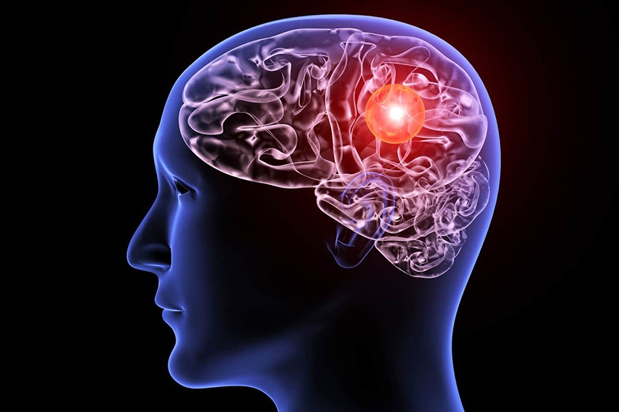
Aneurysm: what is it, symptoms, diagnosis and treatment
An aneurysm is one of the oldest diseases ever recorded by man. According to medical historian Henry Sigerist, the ancient Egyptians already treated it with magical or religious practices, although they never coined a specific term to identify it
We can associate Egyptian treatments with pathology thanks to the accurate description of it in the Ebers Papyrus (dating back to around 1550 B.C.), where it speaks of a vascular lesion to be treated by means of an iron instrument, previously passed over fire.
As for the first treatments, however, we have to wait for the Greek surgeon Antillus (born and lived in the 2nd century AD).
THE RADIO OF RESCUERS AROUND THE WORLD? IT’S RADIOEMS: VISIT ITS BOOTH AT EMERGENCY EXPO
A permanent pathological dilatation, an aneurysm presents itself as a wall bulge that – in most cases – affects the arteries
The vessel wall affected by an aneurysm is weakened to such an extent that the bulging can facilitate bursting and copious bleeding.
Among the most dangerous aneurysms are those affecting the arteries of the brain, a major cause of stroke, or the aorta, which can cause fatal haemorrhaging within minutes.
It is also important to know that, even if an aneurysm does not rupture, it can still impede proper blood circulation and encourage the formation of blood clots or thrombi.
What is an aneurysm and how to recognise it?
An aneurysm is an eversion (or dilation) affecting the wall of a blood vessel, usually an artery; it is formed due to a weakening caused by trauma or pathological alteration.
Aneurysms are often caused by a chronic increase in arterial pressure, but all other pathologies or traumatic events capable of inducing a weakening of the arterial wall can also be responsible for their occurrence.
Some aortic aneurysms can be attributed to hereditary pathologies such as Marfan syndrome, an alteration of the connective tissues that are thus weakened, but age is also one of the causes as – with the passage of time – the vessel walls become less elastic and more prone to dilation.
With regard to aneurysms of an arterial nature (the most common), they appear as a continuous pulsating dilation of the vessel, often associated with degenerative aetiologies such as arteriosclerosis or inflammatory processes due to infectious and/or vascular diseases.
The forms mainly affecting the cerebral arteries are often determined by a congenital or hereditary weakness of the arterial wall (caused by a minor development of the vessel wall).
Unfortunately, the symptoms associated with this condition are particularly sparse and non-specific and do not allow for a prompt diagnosis, which often occurs accidentally while the patient is being checked for other disorders.
In the most unfortunate individuals, the diagnosis is made at the same time as the most serious complication of an aneurysm, namely its rupture.
Patients who are more prone to this risk, due to hereditary causes or a greater susceptibility to aneurysms, should undergo regular check-ups and thus take the necessary preventive measures.
Aneurysm: the causes
The most frequent causes of aneurysm formation are atherosclerosis and hypertension, but there are many other factors responsible for the weakening of the blood vessel wall that can potentially contribute to the onset of the pathology.
Among the most important risk factors are:
- fibromuscular dysplasia
- obesity
- diabetes
- age over 60 (more frequent in males)
- alcoholism
- hypercholesterolaemia
- smoking
- chronic obstructive pulmonary disease
The main causes of aneurysm formation are:
A congenital weakness of the muscular tonaca of the arterial wall including:
- destruction of the elastic or muscular component of the middle tonaca
- genetic predisposition
- production of modified collagen, unable to tolerate pressure or degenerative insults (Marfan syndrome)
- altered balance between metalloproteases – i.e. molecules capable of degrading the components of the extracellular matrix (collagen, elastin, proteoglycans, laminin, etc.) – and their inhibitors.
- Trauma suffered by the blood vessel (prosthesis insertion, thoracic trauma, post-infarct lacerations, etc.).
- Vascular diseases, such as atherosclerosis, vasculitis, syphilis or other infections.
- Infectious diseases, such as syphilis in an advanced stage (typically the third), tuberculosis that can lead to Rasmussen’s aneurysm and infections in the brain that cause infectious intracranial aneurysms.
Types of aneurysm
The various types of aneurysm can be classified according to the site where the pathology is localised, and the blood vessel affected by the bulging and weakening.
An aneurysm can therefore occur:
- In the heart: it affects the aorta, the main artery (thoracic or abdominal aortic aneurysm), and thus involves the large blood vessel that carries arterial blood, rich in oxygen, from the heart to the peripheral vessels.
- In the brain: affects the cerebral arteries (cerebral aneurysm) and consists of the circumscribed dilatation of an intracranial artery (or vein)
- In the arteries of the limbs, affecting the leg at the level of the knee (popliteal artery aneurysm)
- In the visceral arteries, affecting the intestine (mesenteric artery aneurysm) or the spleen (splenic artery aneurysm).
As far as the anatomo-pathological classification is concerned, a distinction is made:
- True aneurysm: characterised by thinning of the elastic lamina of the middle tonaca, which constitutes the wall of the vessel and which may be altered qualitatively or quantitatively.
- Compound aneurysm: consists of a true aneurysm, which over time ruptures the adventitia, i.e. the outermost part of the vessel wall
- False aneurysm: all of the blood vessel’s tonsils are ruptured and the aneurysm wall is formed by the surrounding tissue.
On the basis of the shape, a distinction is made:
- Sacciform aneurysms: they involve short stretches (5-20 cm), for part of the circumference, often occupied by thrombi.
- Navicular aneurysms: they involve short tracts, for the entire circumference.
- Fusiform aneurysms: they affect long stretches (up to 20 cm), and originate following a progressive but gradual dilatation of the entire circumference of the vessel.
- Cylindrical aneurysms: they affect long stretches, the entire circumference of the vessel.
Symptoms vary depending on the site where the pathology is located:
A) Cerebral aneurysm: symptoms may occur if the bulge pushes on an encephalic structure
B) Intact: symptoms may occur in the case of an intact aneurysm, such as
- fatigue
- difficulty of perception
- loss of balance
- aphasia
- double vision
C) Rupture: in the case of a ruptured blood vessel, typical symptoms of subarachnoid haemorrhage may occur
- severe headache
- blindness
- diplopia
- neck pain or stiffness
- pain above or behind the eyes
D) Abdominal aortic aneurysm (usually asymptomatic):
Intact may cause in rare cases
- back pain
- ischaemia of the lower limbs
Rupture:
- rupture manifests with severe hypovolaemic shock that can quickly lead to death.
Renal artery aneurysm:
Intact (facilitates the formation of clots that partially or totally obstruct the artery itself):
- arterial hypertension
- flank pain
- haematuria
- nausea
- vomiting
- acute renal failure (severe cases)
Rupture:
- rupture is manifested by severe hypovolaemic shock which can lead to kidney infarction
How is the aneurysm diagnosed?
An aneurysm cannot be diagnosed in advance unless one undergoes periodic check-ups (especially in cases that are more prone to the possibility of the disease occurring), or unless there is the fortuitous discovery of a visible bulge attributable to the pathology.
In addition to the objective examination and history aimed at searching for risk factors, useful diagnostic tests during the clinical course are
- transesophageal or abdominal ultrasound: this allows the aneurysm to be visualised and to identify the possible presence of a thrombosis. It also makes it possible to verify the evolution of the aneurysm and to check whether it may lead to complications.
- X-ray of the abdomen and thorax (aortic aneurysm): it highlights a large shadow at the level of the lesion and the possible compression of adjacent structures.
- electrocardiogram (in case of aortic involvement)
- magnetic resonance angiography (angio-RM): highlights the vascular district at certain locations
- computed axial tomography angiography (angio-CT, with contrast medium): provides information regarding the extent of the aneurysm, the possibility of a rupture and the possible presence of thrombi that obstruct or prevent normal blood circulation.
The risk of rupture can be assessed on the basis of size, calculated using ultrasound imaging techniques.
Aneurysm: the most effective treatments
Treatment depends mainly on the type, size and location of the aneurysm.
Drug therapy initially involves reducing blood pressure values by administering vasodilators or beta-blockers.
If the aneurysm is small and there are no symptoms, the doctor may recommend regular check-ups, to verify the evolution and to evaluate a possible timely surgical approach.
Should surgery be necessary, several techniques can be employed:
- traditional repair (open): an aneurysm in an accessible area, such as in the abdomen, can be surgically removed and the vessel repaired or replaced with an artificial graft. The prognosis is usually excellent;
- extravascular surgical approach (clipping): allows surgical intervention on the aneurysmal sac to exclude it from circulation;
- endovascular technique (endovascular embolisation): a micro-catheter (very thin tube passing through the blood vessels) is used to reach the site of the aneurysm in order to place a stent. The procedure initiates a coagulation reaction (self-thrombisation) that will strengthen the altered blood vessel wall. This approach is considered the safest, especially in the case of a cerebral aneurysm.
Aneurysm: how to prevent it and effects on daily life
An aneurysm is a very difficult pathology to identify in affected individuals, and often this moment coincides with the bursting of the affected blood vessel and admission to hospital.
In order to prevent the appearance of an aneurysm, it is a good idea to carry out targeted periodic checks, especially for those subjects who are more prone to the onset of this pathology for congenital reasons or due to trauma.
It should also be remembered that obese subjects or smokers are also among those at risk and therefore periodic check-ups are strongly recommended.
Read Also
Emergency Live Even More…Live: Download The New Free App Of Your Newspaper For IOS And Android
Abdominal Aortic Aneurysm: Epidemiology And Diagnosis
Abdominal Aortic Aneurysm: What It Looks Like And How To Treat It
Cerebral Aneurysm: What It Is And How To Treat It
Ruptured Aneurysms: What They Are, How To Treat Them
Pre-Hospital Ultrasound Assessment In Emergencies
Unruptured Brain Aneurysms: How To Diagnose Them, How To Treat Them
Ruptured Brain Aneurysm, Violent Headache Among The Most Frequent Symptoms
Concussion: What It Is, Causes And Symptoms
Ventricular Aneurysm: How To Recognise It?
Ischaemia: What It Is And Why It Causes A Stroke
How Does A Stroke Manifest Itself? Signs To Watch Out For
Treatment Of Urgent Stroke: Changing Guidelines? Interesting Study In The Lancet
Benedikt Syndrome: Causes, Symptoms, Diagnosis And Treatment Of This Stroke
What Is A Positive Cincinnati Prehospital Stroke Scale (CPSS)?
Foreign Accent Syndrome (FAS): The Consequences Of A Stroke Or Severe Head Trauma
Acute Stroke Patient: Cerebrovascular Assessment
Basic Airway Assessment: An Overview
Emergency Stroke Management: Intervention On The Patient
Stroke-Related Emergencies: The Quick Guide


