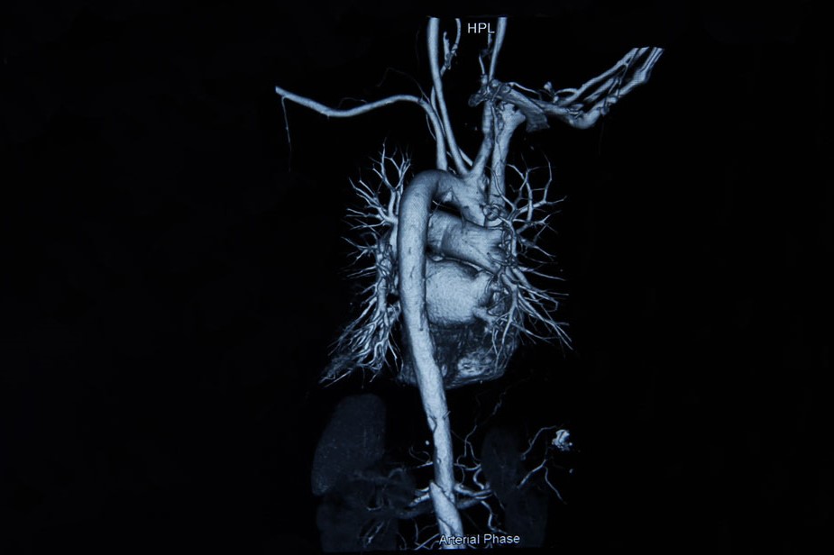
Angiography: what it is and what it is used for
Angiography is the most precise test for assessing the shape and calibre of arteries. It is a technique of radiological visualisation of an artery determined by the introduction of a radiopaque contrast agent
What is angiography used for
Mainly used to diagnose arterial vascular changes in the heart (coronarography), large vessels (aortography) and peripheral thoraco-abdominal or upper or lower limb arteries (arteriography).
Cerebral angiography, since the advent of CT (Computed Axial Tomography) and MRI (Magnetic Resonance Imaging), has lost importance for assessing the exact location and degree of vascularisation of intracranial lesions, but it remains of primary importance for the diagnosis of stenosis, occlusions and aneurysms of extracranial carotid and vertebral arteries or intracranial arteries, as well as congenital anomalies or malformations of cerebral vessels such as AVMs, and thrombosis of the venous sinuses of the dura mater.
Angiography is also a highly sensitive technique in other body districts; it allows precise localisation of pancreatic cell tumours, detection of pulmonary emboli, lesions of the renal vasculature (by integrating CT data), ischaemia or haemorrhage in the gastrointestinal tract or malformations of liver tissue.
This is because the neoformations have abundant and disordered arterial vascular systems that are easily detected by this test.
How angiography is performed
Angiography is performed by cannulating, using special catheters, the arteries of the organ to be examined, then introducing the contrast medium (iodine-based) and recording the X-ray images at a rate of approximately 3-6 sec.; the contrast medium is then eliminated via the kidney.
Angiography and possible side effects
Transient side effects such as feelings of heat, drop in blood pressure, nausea may occur during the test.
True allergic reactions to the contrast medium (bronchospasm, oedema of the larynx, urticaria) are rare, while more severe complications such as cardiac arrhythmias, anaphylactic reactions and shock are considered exceptional but possible (in the order of 4 per 100,000 individuals).
In approximately 2% of cases, angiography may result in mild renal failure.
Contraindications to the test are previous allergic reactions to iodinated contrast medium and renal insufficiency.
Since the occurrence of these side effects cannot be predicted, it is essential to perform the test in centres equipped with resuscitative equipment and an anaesthetist, and only if strictly necessary.
Conventional angiography has recently been refined for some indications using the digital subtraction technique, which improves image quality and makes it possible to use smaller quantities of contrast medium and smaller calibre catheters.
Read Also
Emergency Live Even More…Live: Download The New Free App Of Your Newspaper For IOS And Android
What Is ECO Fusion (Fusion Ultrasound) Used For?
What Is Magnetic Resonance Angiography (MRA)?
What Is Retinal Fluorangiography And What Are The Risks?
Nuclear Magnetic Resonance (NMR): When To Do It?
Medical Thermography: What Is It For?
Rheumatic Diseases: The Role Of Total Body MRI In Diagnosis
Positron Emission Tomography (PET): What It Is, How It Works And What It Is Used For
Single Photon Emission Computed Tomography (SPECT): What It Is And When To Perform It
Instrumental Examinations: What Is The Colour Doppler Echocardiogram?
Coronarography, What Is This Examination?
CT, MRI And PET Scans: What Are They For?
MRI, Magnetic Resonance Imaging Of The Heart: What Is It And Why Is It Important?
Urethrocistoscopy: What It Is And How Transurethral Cystoscopy Is Performed
What Is Echocolordoppler Of The Supra-Aortic Trunks (Carotids)?
Surgery: Neuronavigation And Monitoring Of Brain Function
Robotic Surgery: Benefits And Risks
Refractive Surgery: What Is It For, How Is It Performed And What To Do?


