Asbestosis, mesothelioma and pleural effusion: causes, symptoms, treatment
Asbestosis is a type of pneumoconiosis, i.e. a lung disease caused by the inhalation of dust, for example at work, and in fact asbestosis is a typical occupational respiratory disease
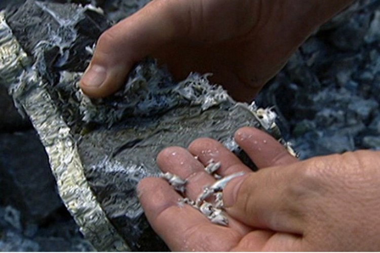
Asbestosis, in particular, is a diffuse interstitial pneumoconiosis that originates from the prolonged inhalation of asbestos dust (silicate mineral fibres of different chemical composition) during the extraction, milling, industrial processing, application (e.g. for insulation) or removal of asbestos products.
The risk of developing asbestosis, lung cancer and mesothelioma depends on cumulative exposure to asbestos fibres over a lifetime
It appears that asbestos promotes but does not initiate carcinogenesis.
The incidence of lung cancer is increased in smokers with asbestosis, with a dose-dependent relationship.
Whether there is an increased risk in non-smokers is doubtful, but, if present, it is considered minimal.
The probability of developing lung cancer is significantly increased in persons who are exposed to asbestos and are heavy smokers, especially if they smoke > 1 packet/day.
Various related diseases, such as mesotheliomas and pleural effusions, can be associated with asbestosis, in addition to lung cancer.
Asbestosis: Malignant Pleural and Peritoneal Mesotheliomas
These are rare tumours of mesothelial tissue associated with exposure to asbestos.
Exposure has consistently occurred 15-40 years previously and may have been relatively short-lived (i.e., 12 months), but intense.
Mesothelioma is usually associated with exposure to crocidolite, one of the four major commercial fibres.
Amosite also often causes mesothelioma, but the tumour is very rare in individuals exposed to chrysolite and anthophyllite.
Studies suggest that the tumour in a person exposed to chrysolite usually arises from chrysolite deposits that are contaminated by tremolite, a non-commercial amphibole form of asbestos.
Malignant pleural mesotheliomas, although rare, are more frequent than benign ones.
The malignant tumour is diffuse, extensively infiltrates the pleura and is always associated with pleural effusion.
The fluid may be viscous due to the high concentration of hyaluronic acid.
Benign pleural plaques and pleural effusions may develop after exposure to asbestos; however, benign pleural mesotheliomas are not related to asbestos exposure.
Pleural effusion from asbestosis
People exposed to asbestos rarely develop a pleural effusion 5-20 years after exposure.
The effusion may follow a short exposure but more often it follows intermediate exposures (i.e. up to 10-15 years).
The mechanism is unknown, but it is assumed that the fibres move from the lungs to the pleura inducing an inflammatory response.
In most people, the effusions resolve after 3-6 months; 20% develop diffuse pleural fibrosis.
A few individuals may develop malignant mesothelioma many years later, but there is no evidence of an increased incidence of mesothelioma among those who have had a pleural effusion.
Pathological Anatomy and Pathophysiology
Individual asbestos fibres can be inhaled deeply into the lung parenchyma and, once deposited and retained, cause the development of diffuse alveolar and interstitial fibrosis.
Asbestosis leads to a reduction in volume, compliance (with increased stiffness) and gas exchange.
The asbestos fibres in the lung tissue may or may not be coated with an iron-protein complex.
Once the fibres are coated (ferruginous or asbestos bodies), they are considered to become harmless.
In the absence of signs of fibrosis in the lungs, the presence of fibres in lung tissue only indicates asbestos exposure, not disease.
Occasionally, other fibres, e.g. talc coated with an iron-containing protein, may mimic asbestos fibres.
Asbestosis, symptoms and signs
The patient characteristically experiences the insidious onset of exertional dyspnoea and reduced exercise tolerance.
Symptoms of airway pathology (coughing, sputum and wheezing) are uncommon, but may occur in heavy smokers with associated chronic bronchitis.
The chest X-ray reveals small irregular or linear opacities that are diffusely distributed, usually more evident in the lower lung fields.
Often only minimal changes are visible on X-ray and these are often confused with those related to other conditions.
Diffuse or localised pleural thickening, associated or not with parenchymal pathology, may also be evident.
The disease progresses (but only for 1-5 years) in about 5-12% of patients after cessation of exposure.
Symptomatology and functional impairment become more severe as the rx category worsens. This eventually leads to respiratory failure with marked impairment of oxygenation.
Localised pleural plaques do not impair function, although diffuse fibrosis of the pleura, as occurs after a pleural effusion, is usually associated with severe restrictive impairment.
Mesotheliomas associated with asbestos exposure are almost always fatal within 2-4 years of diagnosis.
They spread locally by extension and may metastasise diffusely.
A pleural effusion with chest pain is often present.
Diagnosis
The diagnosis of asbestosis requires a history of occupational exposure and x-ray, clinical and functional findings of restrictive damage and reduced diffuse pulmonary fibrosis.
Histological confirmation is almost never necessary or indicated.
Although the diagnosis of bronchogenic carcinoma can be quickly made, there are serious medico-legal problems in establishing a cause-and-effect relationship with exposure to asbestos fibres in the individual patient, especially if he or she is a cigarette smoker.
Only when asbestosis is clearly demonstrated can it be assumed that asbestos exposure played a role.
It is more difficult to diagnose mesothelioma, which can only be confirmed at biopsy or autopsy.
Prophylaxis and treatment
Asbestosis is preventable, especially with effective removal of dust from the work environment.
The sharp decline in asbestos exposure has reduced the incidence of asbestosis and further advances in industrial hygiene are likely to eradicate it.
The most effective prophylaxis against lung cancer can be implemented by the worker, i.e. avoiding continuous exposure and, above all, refraining from smoking cigarettes.
Since short exposure to asbestos (at least 6 months to 2 years) but generally intense can lead to the development of mesothelioma, its prevention cannot be predicted with certainty, but its incidence will be much lower now that crocidolite is no longer used in North America and most of Europe.
There is no specific therapy for asbestosis or mesothelioma; treatment is symptomatic.
Other occupational respiratory diseases
Other frequent occupational respiratory diseases that might interest you are:
- silicosis;
- coal workers’ pneumoconiosis;
- berylliosis;
- hypersensitivity pneumonitis;
- occupational asthma;
- byssinosis;
- diseases caused by irritant gases and other chemicals;
- sick building syndrome.
Read Also
Emergency Live Even More…Live: Download The New Free App Of Your Newspaper For IOS And Android
Bronchial Asthma: Symptoms And Treatment
Bronchitis: Symptoms And Treatment
Bronchiolitis: Symptoms, Diagnosis, Treatment
Extrinsic, Intrinsic, Occupational, Stable Bronchial Asthma: Causes, Symptoms, Treatment
Chest Pain In Children: How To Assess It, What Causes It
Bronchoscopy: Ambu Set New Standards For Single-Use Endoscope
What Is Chronic Obstructive Pulmonary Disease (COPD)?
Respiratory Syncytial Virus (RSV): How We Protect Our Children
Respiratory Syncytial Virus (RSV), 5 Tips For Parents
Infants’ Syncytial Virus, Italian Paediatricians: ‘Gone With Covid, But It Will Come Back’
Respiratory Syncytial Virus: A Potential Role For Ibuprofen In Older Adults’ Immunity To RSV
Neonatal Respiratory Distress: Factors To Take Into Account
Stress And Distress During Pregnancy: How To Protect Both Mother And Child
Respiratory Distress: What Are The Signs Of Respiratory Distress In Newborns?
Respiratory Distress Syndrome (ARDS): Therapy, Mechanical Ventilation, Monitoring
Bronchiolitis: Symptoms, Diagnosis, Treatment
Chest Pain In Children: How To Assess It, What Causes It
Bronchoscopy: Ambu Set New Standards For Single-Use Endoscope
Bronchiolitis In Paediatric Age: The Respiratory Syncytial Virus (VRS)
Pulmonary Emphysema: Causes, Symptoms, Diagnosis, Tests, Treatment
Bronchiolitis In Infants: Symptoms
Fluids And Electrolytes, Acid-Base Balance: An Overview
Ventilatory Failure (Hypercapnia): Causes, Symptoms, Diagnosis, Treatment
What Is Hypercapnia And How Does It Affect Patient Intervention?
Symptoms Of Asthma Attack And First Aid To Sufferers
Occupational Asthma: Causes, Symptoms, Diagnosis And Treatment


