
Aslanger Pattern: Another OMI?
In April 2020, Aslanger et al identified a specific ECG pattern concerning for acute inferior occlusion MI (OMI) in patients with concomitant multi-vessel disease, that does not display contiguous ST-segment elevation or fulfil STEMI criteria
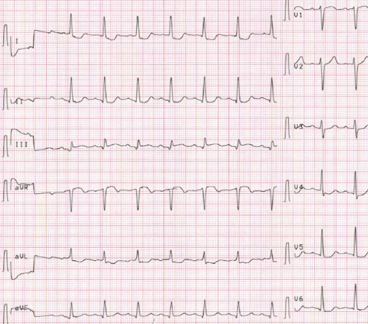
The Aslanger Pattern
The publishers reviewed ECG and angiography findings from 1000 NSTEMI, 1000 control (no myocardial infarction), as well as inferior STEMI patients presenting during the same time period.
The Aslanger Pattern was observed in 6.3% of NSTEMI patients and found to be a predictor of larger infarct size and higher mortality.
ECG Criteria
1) Inferior STE isolated to lead III
2) Concomitant ST depression in any of V4-V6, with a positive/terminally positive T-wave
3) ST segment in V1 > V2
Aslanger pattern 2020
(1) STE in III but not in any other inferior lead,
(2) ST depression in any of leads V4 to 6 (but not in V2) with a positive (at least terminally positive) T-wave,
(3) ST in lead V1 higher than ST in V2.
Why is there not contiguous ST elevation?
- In cases of limited inferior wall injury, the ST vector of inferior MI localises the area of infarction and is typically directed inferiorly and rightwards (yellow arrow)
- The ST vector of subendocardial ischaemia does not localise to the ischaemia and regardless of involved coronary region directs to lead aVR (blue arrow)
- The resultant average ST vector directs rightwards, causing ST elevation only in lead III and aVR
Aslanger pattern, clinical significance
Concurrent multi-vessel disease predisposes these patients to poor outcomes if there is delayed time to emergent reperfusion, and prompt recognition of this potential OMI should improve outcomes
Identification of the culprit lesion at the time of angiography may be difficult if there is multiple critical stenoses, and this pattern would guide lesions supplying the inferior wall to be opened first
Limitations
- The pattern was found to be present in 0.5% of patients without acute MI, which may be a result of chronic change from a previous ischaemic insult
- Acute inferior MI in the presence of previous infarctions may also change the overall orientation of the lesion vector causing a similiar pattern
- This is an isolated, retrospective study and warrants a further analysis as a predictor of occlusion MI that would be responsive to emergent reperfusion therapy
Associated Persons
Emre Aslanger; Yeditepe University Hospital, Department of Cardiology, Istanbul, Turkey
References
Historical references
Aslanger E, Yıldırımtürk Ö, Şimşek B, Sungur A, Türer Cabbar A, Bozbeyoğlu E, Karabay CY, Smith SW, Değertekin M. A new electrocardiographic pattern indicating inferior myocardial infarction. J Electrocardiol. 2020 Jul-Aug;61:41-46.
Aslanger EK, Smith SW. Response to: “A new electrocardiographic pattern indicating inferior myocardial infarction”. J Electrocardiol. 2020 Nov 18
Eponymous term review
Fiol M, Carrillo A, Cygankiewicz I, Velasco J, Riera M, Bayés-Genis A, Gómez A, Peral V, Bethencourt A, Goldwasser D, Molina F, Bayés de Luna A. A new electrocardiographic algorithm to locate the occlusion in left anterior descending coronary artery. Clin Cardiol. 2009 Nov;32(11):E1-6
Bozbeyoğlu E, Aslanger E, Yıldırımtürk Ö, Şimşek B, Hünük B, Karabay CY, Kozan Ö, Değertekin M. The established electrocardiographic classification of anterior wall myocardial infarction misguides clinicians in terms of infarct location, extent and prognosis. Ann Noninvasive Electrocardiol. 2019 May;24(3):e12628
Turgay Yildirim Ö, Çanakçı ME. The new ECG pattern for inferior myocardial infarction. J Electrocardiol. 2020 Nov-Dec;63:64
Smith SW. Subtle ECG Findings of Left Anterior Descending Artery (LAD) Occlusion — LAD Occlusion MI (OMI). Vimeo 2020
Smith SW. A 58 year old collapses in the hot sun Dr Smith’s ECG Blog 2020
Smith SW. ECG with Aslanger’s Pattern. CT Pulmonary Angiogram Reveals LAD Ischemia (Septal Transmural). But this is not Contradictory. Dr Smith’s ECG Blog 2021
Buttner R. OMI: Replacing the STEMI misnomer. LITFL 2021
Read Also
Emergency Live Even More…Live: Download The New Free App Of Your Newspaper For IOS And Android
ECG: Waveform Analysis In The Electrocardiogram
What Is An ECG And When To Do An Electrocardiogram
ST-Elevation Myocardial Infarction: What Is A STEMI?
ECG First Principles From Handwritten Tutorial Video
ECG Criteria, 3 Simple Rules From Ken Grauer – ECG Recognize VT

Defibrillator: What It Is, How It Works, Price, Voltage, Manual And External
The Patient’s ECG: How To Read An Electrocardiogram In A Simple Way
ECG: What P, T, U Waves, The QRS Complex And The ST Segment Indicate
Endocavitary Electrophysiological Study: What Does This Examination Consist Of?
Head Up Tilt Test, How The Test That Investigates The Causes Of Vagal Syncope Works
Stress Electrocardiogram (ECG): An Overview Of The Test
What Is Ischaemic Heart Disease And Possible Treatments
Percutaneous Transluminal Coronary Angioplasty (PTCA): What Is It?
Ischaemic Heart Disease: What Is It?
EMS: Pediatric SVT (Supraventricular Tachycardia) Vs Sinus Tachycardia
Paediatric Toxicological Emergencies: Medical Intervention In Cases Of Paediatric Poisoning
Valvulopathies: Examining Heart Valve Problems
What Is The Difference Between Pacemaker And Subcutaneous Defibrillator?
Heart Disease: What Is Cardiomyopathy?
Inflammations Of The Heart: Myocarditis, Infective Endocarditis And Pericarditis
Heart Murmurs: What It Is And When To Be Concerned
Clinical Review: Acute Respiratory Distress Syndrome
Botallo’s Ductus Arteriosus: Interventional Therapy
Heart Valve Diseases: An Overview
Cardiomyopathies: Types, Diagnosis And Treatment
First Aid And Emergency Interventions: Syncope
Tilt Test: What Does This Test Consist Of?
Cardiac Syncope: What It Is, How It Is Diagnosed And Who It Affects
New Epilepsy Warning Device Could Save Thousands Of Lives
Understanding Seizures And Epilepsy
First Aid And Epilepsy: How To Recognise A Seizure And Help A Patient
Neurology, Difference Between Epilepsy And Syncope
Positive And Negative Lasègue Sign In Semeiotics
Wasserman’s Sign (Inverse Lasègue) Positive In Semeiotics
Positive And Negative Kernig’s Sign: Semeiotics In Meningitis
Lithotomy Position: What It Is, When It Is Used And What Advantages It Brings To Patient Care
Trendelenburg (Anti-Shock) Position: What It Is And When It Is Recommended
Prone, Supine, Lateral Decubitus: Meaning, Position And Injuries
Stretchers In The UK: Which Are The Most Used?
Does The Recovery Position In First Aid Actually Work?
Reverse Trendelenburg Position: What It Is And When It Is Recommended
Drug Therapy For Typical Arrhythmias In Emergency Patients
Canadian Syncope Risk Score – In Case Of Syncope, Patients Are Really In Danger Or Not?