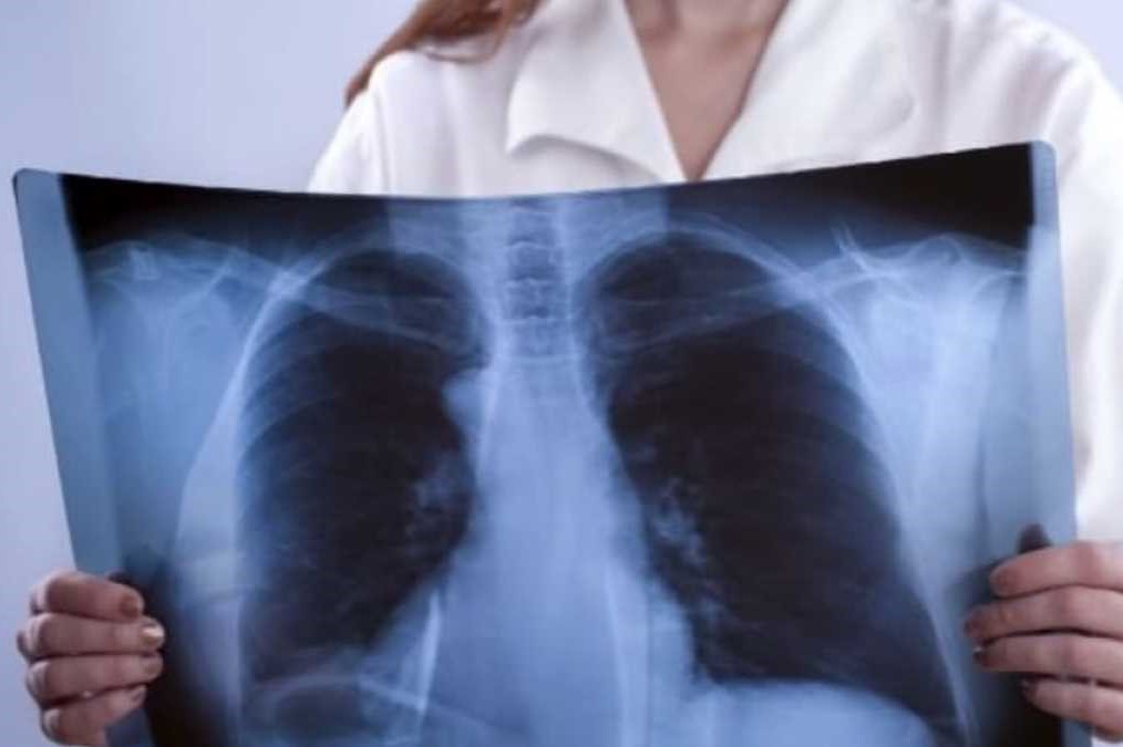
Atelectasis: symptoms and causes of collapsed lung areas
The term atelectasis refers to the collapse of one or more areas of the lung associated with a loss of volume. Under normal conditions, when one inhales, the lungs fill with air
This air travels to the alveoli, the small cavities located at the end of the bronchioles that are responsible for allowing gas exchange between air and blood.
In the case of atelectasis, these small air sacs deflate and cannot inflate properly and/or absorb enough air and oxygen.
If the disease affects a large enough area, the blood may not receive enough oxygen, which can trigger various health problems.
Generally, it is not life-threatening, but in some cases it must be treated quickly.
Atelectasis: what it is
Atelectasis is one of the most common respiratory complications after surgery.
It is also a possible complication of other respiratory problems, including cystic fibrosis, lung tumours, chest lesions, fluid in the lungs and respiratory weakness.
Atelectasis can make breathing difficult, particularly if one already suffers from lung disease.
Treatment depends on the cause and severity of the collapse.
Pulmonary atelectasis, symptoms
What are the signs and symptoms? If atelectasis only affects a small area of the lungs, the person may not even have any symptoms.
But if it affects larger areas, the lungs cannot fill with enough air and the oxygen level in the blood may decrease.
When this happens, annoying and unpleasant symptoms may occur, including:
- difficulty breathing (shortness of breath; rapid, shallow breathing; wheezing);
- increased heart rate;
- coughing;
- chest pain;
- bluish discolouration of the skin and lips.
If you experience these symptoms and have difficulty breathing, you should consult your doctor for diagnosis and treatment.
Keep in mind that other conditions, including asthma and emphysema, can also cause chest pain and breathing problems.
Why a lung can collapse
Atelectasis can be triggered by many factors: potentially, any condition that makes it difficult to take deep breaths or cough can lead to a collapsed lung.
Atelectasis can result from airway obstruction (called obstructive atelectasis) or from pressure from outside the lung (non-obstructive atelectasis).
The most common reason for people to develop this disease is surgery.
It must be known that anaesthesia can affect the patient’s ability to breathe normally or cough as it changes the normal breathing pattern and affects lung gas exchange.
All this can cause the air sacs (alveoli) to deflate.
In addition, the pain that is often experienced following surgery may make deep breathing painful: as a result, one may be inclined to adopt continuous shallow breathing, which may favour the development of the disease.
This explains why almost everyone who has undergone major surgery develops a more or less severe form of atelectasis.
Other possible causes of this pathology are:
- thoracic trauma, e.g. a fall or a car accident, which prevent one from taking deep breaths (due to pain), which can cause compression of the lungs;
- pressure at the level of the chest: pressure exerted on the lungs, which may depend on a tumour mass outside the bronchus, on a tumour inside the bronchus, which causes airway obstruction. In fact, if air cannot get past the blockage present, the affected part of the lung may collapse;
- accumulation of mucus in the airways, which may cause a blockage in the airflow. This event commonly occurs during and after surgery because coughing is not possible in such cases. In addition, drugs administered during surgery cause people to breathe less deeply, so normal secretions collect in the airways. Suctioning the lungs during surgery helps to clear them, but sometimes it is not enough. Mucus plugs are also common in children, people with cystic fibrosis and during severe asthma attacks;
- inhalation of small objects, such as a peanut, the cap of a biro, a small toy, which prevent air from flowing freely;
- other lung diseases, such as pneumonia, pleural effusions (fluid around the lungs) and respiratory distress syndrome (RDS).
Atelectasis is not to be confused with pneumothorax, another condition that commonly causes a collapsed lung.
It is the presence of air between the lung and chest wall.
Atelectasis, the risk factors
Factors that increase the likelihood of developing this disease include:
- advanced age
- any condition that makes swallowing difficult;
- bed confinement with rare changes of position;
- lung disease, such as asthma, COPD, bronchiectasis or cystic fibrosis;
- recent abdominal or thoracic surgery;
- recent general anaesthesia;
- weak respiratory muscles due to muscular dystrophy, spinal cord injury or another neuromuscular condition;
- use of drugs that may cause shallow breathing;
- pain or injuries that may make it painful to cough or cause shallow breathing, including stomach pain or rib fracture;
- cigarette smoking.
What is involved in atelectasis
A small area of atelectasis, especially in an adult, is usually curable.
However, one should be aware that this disease can give rise to the following complications
- a low level of oxygen in the blood (hypoxemia). Atelectasis makes it more difficult for the lungs to carry oxygen to the air sacs (alveoli) and thus to the rest of the body;
- pneumonia: the risk of pneumonia continues until the atelectasis disappears. This is because the presence of mucus in a collapsed lung can lead to infection;
- respiratory failure: the loss of a lobe or an entire lung, particularly in an infant or in people with lung disease, can be life-threatening.
Prevention of post-surgery atelectasis
Some research suggests that performing deep breathing exercises and muscle training may reduce the risk of developing atelectasis after surgery.
In addition, many patients in hospital are given a device called an incentive spirometer that can encourage them to take deep breaths, thus preventing and treating atelectasis.
If you smoke, you can reduce your risk of developing the condition by stopping smoking before any operation.
Atelectasis in children is often caused by an airway blockage.
In such cases, to reduce the risk of atelectasis, keep small objects out of reach of children.
WOULD YOU LIKE TO GET TO KNOW RADIOEMS? VISIT THE RESCUE RADIO BOOTH AT EMERGENCY EXPO
Read Also
Emergency Live Even More…Live: Download The New Free App Of Your Newspaper For IOS And Android
Benefits And Risks Of Prehospital Drug Assisted Airway Management (DAAM)
Blind Insertion Airway Devices (BIAD’s)
Pulmonological Examination, What Is It And What Is It For? What Does The Pulmonologist Do?
Oxygen-Ozone Therapy: For Which Pathologies Is It Indicated?
Hyperbaric Oxygen In The Wound Healing Process
Venous Thrombosis: From Symptoms To New Drugs
What Is Intravenous Cannulation (IV)? The 15 Steps Of The Procedure
Nasal Cannula For Oxygen Therapy: What It Is, How It Is Made, When To Use It
Pulmonary Emphysema: Causes, Symptoms, Diagnosis, Tests, Treatment
Extrinsic, Intrinsic, Occupational, Stable Bronchial Asthma: Causes, Symptoms, Treatment
A Guide To Chronic Obstructive Pulmonary Disease COPD
Bronchiectasis: What Are They And What Are The Symptoms
Bronchiectasis: How To Recognise And Treat It
Pulmonary Vasculitis: What It Is, Causes And Symptoms
Bronchiolitis: Symptoms, Diagnosis, Treatment
Chest Pain In Children: How To Assess It, What Causes It
Bronchoscopy: Ambu Set New Standards For Single-Use Endoscope
What Is Chronic Obstructive Pulmonary Disease (COPD)?


