
Atrial fibrillation: causes, symptoms and treatment
Atrial fibrillation is the most common form of cardiac arrhythmia and is characterised by the presence of disorganised, very rapid and mechanically ineffective atrial electrical activity (the atrium does not contract in a rhythmic and coordinated manner with the activity of the ventricles)
The incidence of this arrhythmia in the population is around 1%, but increases with age, reaching 6% in people over 60.
It can be permanent (stable over time), paroxysmal (short episodes that resolve on their own) or persistent (prolonged episodes that require medical intervention to stop).
Causes of atrial fibrillation
Atrial fibrillation can be the consequence of cardiac problems such as valvulopathies (especially mitral and aortic stenosis or insufficiency), heart muscle diseases (myocarditis, cardiomyopathies, chronic heart failure), congenital heart disease, myocardial infarction or other disorders such as hypertension, thyroid disease, pulmonary embolism, hydro-electrolyte imbalance.
QUALITY AED? VISIT THE ZOLL BOOTH AT EMERGENCY EXPO
It can also be due to medication (anti-inflammatory drug abuse) or drugs.
A frequent cause is ethyl abuse, so much so that many episodes occur at weekends, when alcohol consumption is generally highest.
Other causes are obesity, states of stress and metabolic syndrome.
Arrhythmia can also occur immediately after heart surgery, due to alterations in electrolytes and the ‘stress’ suffered by the heart.
Sometimes, particularly in young people but also in older age groups, it is not possible to identify a precise cause (idiopathic atrial fibrillation).
Atrial fibrillation: the consequences
During arrhythmia, there is a lack of effective and regular atrial contraction.
The atrial chambers are practically immobile and progressively dilate.
Rapid atrial electrical activity (up to > 400/min) is conducted as usual to the ventricles via the atrio-ventricular node, which filters and reduces the frequency of the impulses passing through it.
The ventricular rate is still high, usually around 150-160 beats per minute in the absence of therapy, with instantaneous rates that can exceed 200/min.
The consequences of the loss of the mechanical function of the atrium and thus its contribution to filling the ventricle vary from subject to subject.
In the absence of organic heart disease, a paroxysmal atrial fibrillation of short duration (hours) is generally well tolerated, without any haemodynamic repercussions (blood pressure remains normal and the only symptom felt by the subject may be an annoying sense of palpitation).
In the case of associated organic heart disease, the onset of this arrhythmia may, on the other hand, lead to a more rapid deterioration of cardiac compensation.
Thus, while in some cases the arrhythmia does not affect the quantity and quality of life, in others there may be a worsening of the prognosis and quality of life.
The risks of atrial fibrillation
As a result of the loss of contractile capacity and enlargement of the atria, the blood velocity in the right and left atrial chambers decreases significantly.
This slowing down can lead to the formation of aggregates of blood cells and proteins (thrombi).
Thrombi mainly form in small eversions of the atria called auricles, which represent an embryonic and ancestral remnant of the heart.
If these thrombi remain at the atrial level, they do not cause any problems, but often parts of them (emboli) suddenly enter the circulation and end up in the lungs (pulmonary embolism), the brain (stroke), the heart (myocardial infarction), the intestinal vessels (intestinal infarcts) or the kidneys (renal infarcts), the limbs (acute occlusion with ischaemia of the affected limb) or any other organ.
Symptoms of atrial fibrillation
In the presence of atrial fibrillation, symptoms are extremely variable. Some patients experience no discomfort at all, others feel a rapid and sudden heartbeat.
Still others feel they have difficulty breathing (dyspnoea), feel tired or experience chest pain.
In patients who already had other heart problems, the onset of atrial fibrillation can even lead to heart failure, pulmonary oedema and require urgent in-patient treatment.
How arrhythmia is recognised
Recognising arrhythmia is sometimes very easy. If the symptoms described above are present, simply check the pulse.
If an irregular and tachycardic (racing) pulse is found, the diagnosis of atrial fibrillation is very likely.
Sometimes, however, the patient does not complain of any symptoms, and the arrhythmia may therefore be recognised by chance by performing an electrocardiogram for other reasons or during a hospitalisation for a complication of the arrhythmia (stroke).
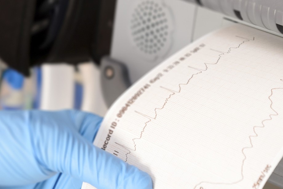
For confirmation of the clinical suspicion, however, it is essential to perform an electrocardiogram or to use prolonged monitoring with a HOLTER ECG or a single-derivative hand-held ECG; in selected cases, it is even necessary to insert a small device called a LOOP RECORDER under the skin, which makes it possible to detect occult episodes of atrial fibrillation as a cause of syncope or neurological events with an undetermined cause.
Evolution of the pathology
Atrial fibrillation may be paroxysmal with occasional episodes, often resolving spontaneously, but sometimes it requires treatment with drugs or other means to restore normal rhythm (sinus rhythm).
In the presence of infarction symptoms and fibrillation symptoms that are particularly relevant in terms of haemodynamic impairment (e.g. hypotension), restoration of rhythm may be urgent.
In the absence of disabling symptoms and in particular clinical conditions, the persistence of atrial fibrillation may also be acceptable (‘chronic’ atrial fibrillation).
What to do in the event of arrhythmia
Within 48 hours of the onset of arrhythmia, sinus rhythm can be restored without delay, because the probability of atrial thrombus formation is very low.
It is therefore advisable to go to the emergency room as soon as possible, at least in cases where the onset of arrhythmia is recognisable by symptoms.
If we cannot be certain of the time of onset of the arrhythmia, or if we are certain that the arrhythmia has been present for more than 48 hours, it is necessary to institute appropriate anticoagulant therapy for 3-4 weeks before attempting to restore sinus rhythm, which prolongs and complicates treatment.
Atrial fibrillation: treatment
The first step is to control the heart rate, which can be slowed down with drugs that reduce the frequency of conduction of electrical impulses to the ventricle, while atrial fibrillation persists.
It is then important to assess the duration of the arrhythmia and the patient’s symptoms for the risks described above.
If atrial fibrillation has been present for less than 48 hours, immediate restoration of sinus rhythm may be considered; otherwise, a period of 3-4 weeks of adequate oral anticoagulation is required before attempting to stop the arrhythmia.
Restoration of the rhythm can be performed by administering anti-arrhythmic drugs (pharmacological cardioversion), or by resorting to electrical therapy (electrical cardioversion).
In some cases it is necessary to restore sinus rhythm urgently (in patients with angina, pulmonary oedema or cardiogenic shock).
Cardioversion causes resynchronisation of atrial electrical and mechanical activity
Often after cardioversion, atrial fibrillation recurs and daily antiarrhythmic drugs must be taken to maintain sinus rhythm.
The restoration of sinus rhythm does not always correspond to an immediate restoration of atrial contraction.
It is therefore necessary to continue oral anticoagulation for at least 4 weeks after the end of the arrhythmia and often even longer, not infrequently forever.
However, this decision must be individualised, bearing in mind each patient’s clinical condition and risk factors.
In order to avoid the pre-cardioversion anticoagulation period, a transesophageal echocardiogram is increasingly being performed, which makes it possible to visualise any thrombi at the level of the heart chambers (which cannot be fully visualised with transthoracic ultrasound).
If the examination is normal, cardioversion can be carried out directly.
This reduces the overall duration of atrial fibrillation and increases the success rate of the procedure.
The arrhythmia in fact tends to self-maintain: the longer it lasts, the more difficult it is to interrupt it.
If the restoration of sinus rhythm fails, or if it is not deemed indicated, the decision is made to ‘chronicise’ the atrial fibrillation, i.e. to leave the patient in atrial fibrillation, controlling the heart rate with the appropriate drugs and administering oral anticoagulant therapy at the same time.

If oral anticoagulant therapy is deemed too risky, antiplatelet drugs will be used instead.
The results of a number of international studies have provided much relevant information regarding the clinical management of this arrhythmia.
There is no difference in terms of duration and quality of life between a treatment strategy that seeks to restore and maintain sinus rhythm and one in which atrial fibrillation is allowed to become chronic, maintaining good anticoagulation and controlling only the heart rate.
In selected cases, it is also possible to resort to other treatments such as radiofrequency ablation, which produces ‘burns’ on the inside of the heart, isolating the points from which the arrhythmia originates, especially at the level of the outlet of the four pulmonary veins in the left atrium.
This method, however, although very promising, is not yet capable of resolving all arrhythmias because, as already described, the conditions favouring and triggering them are many and heterogeneous.
The indication for ablation and the probability of success of the method (between 50 and 80%) is higher in younger subjects in whom the arrhythmia is paroxysmal, the atrium is not dilated and there are no comorbidities or concomitant associated cardiac pathologies.
If no other treatment is possible, because drugs prove ineffective or are not tolerated, ablation of the atrioventricular node, i.e. destruction of the electrical impulse conduction pathway from the atrium to the ventricle, may sometimes be used.
In this case, however, the implantation of a pace-maker is necessary to effectively suppress the cardiac electrical activity.
Side effects must always be taken into account in the choice of therapy: oral anticoagulant therapy can cause bleeding, therapy with anti-arrhythmic drugs can even cause dangerous ventricular arrhythmias; invasive methods (ablation) are also not without risk (pulmonary vein stenosis or haemopericardium).
When a patient in atrial fibrillation cannot take any anticoagulant due to severe bleeding conditions or concomitant risky pathologies in the history (e.g. oesophageal varices, ulcerative rectocolitis, previous intracranial haemorrhagic stroke), the left atrial auricle can be closed with specific devices in order to prevent the formation of clots in the atrium.
Anticoagulants in atrial fibrillation
Anticoagulants are essential in the prophylaxis of cardioembolic stroke and the prevention of recurrences.
Historically, anticoagulants termed vitamin K antagonists (dicumarolics: warfarin and acenocoumarol) have been used, with a therapeutic range measured by a blood test called INR, i.e. prothrombin time, which eliminates the variability of results obtained in different laboratories.
This value is generally used for people taking anticoagulant drugs, in which case it should be between 2.0 and 3.0.
In the absence of particular problems, however, values between 0.9 and 1.3 are considered normal.
The limitation of using dicumarolics concerns the need for frequent blood samples to check INR values and thus to modify the dosage of the drug, and the interaction with many foods containing Vitamin K (especially green leafy vegetables) that reduce its effect, and many interactions with other drugs that modify its bioavailability.
It must be remembered that the use of antiplatelets instead of anticoagulants does not significantly reduce the risk of stroke at a slightly lower risk of bleeding.
In recent years, new oral anticoagulants (NAO) with different dosages have arrived that have demonstrated an efficacy and safety profile equivalent to and even superior to Warfarin with the added advantage of not requiring any periodic blood sampling except for at least six-monthly renal function checks.
Some NAOs also have a specific antidote drug to antagonise their effect and limit episodes of severe acute bleeding.
The prescription of NAOs is possible after having completed a treatment plan that includes the calculation of the ischaemic and haemorrhagic risk profile with specific scores.
Prevention of atrial fibrillation
Effective prevention of atrial fibrillation is only possible in certain cases.
In patients with valvulopathies or certain congenital heart diseases, if indicated, surgery can be performed before the atria dilate excessively.
Atrial dilatation is in fact a factor that favours the onset of arrhythmia.
Adequate blood pressure control as well as avoiding excessive alcohol consumption are useful measures to prevent atrial fibrillation.
Therefore, the choice of pharmacological therapy with anticoagulants and antiarrhythmics must be planned and monitored over time by the cardiologist.
The therapeutic choice with electrical cardioversion or ablation, left auricular occlusion are therapies that must be individualised for each case.
Read Also:
Emergency Live Even More…Live: Download The New Free App Of Your Newspaper For IOS And Android
Cardiac Rhythm Disturbance Emergencies: The Experience Of US Rescuers
Prenatal Pathologies, Congenital Heart Defects: Pulmonary Atresia
Management Of Cardiac Arrest Emergencies
Palpitations: What Causes Them And What To Do
The J-Curve Theory In High Blood Pressure: A Really Dangerous Curve
Why Children Should Learn CPR: Cardiopulmonary Resuscitation At School Age
What Is The Difference Between Adult And Infant CPR
Long QT Syndrome: Causes, Diagnosis, Values, Treatment, Medication
What Is Takotsubo Cardiomyopathy (Broken Heart Syndrome)?
The Patient’s ECG: How To Read An Electrocardiogram In A Simple Way
Stress Exercise Test Inducing Ventricular Arrhythmias In LQT Interval Individuals
CPR And Neonatology: Cardiopulmonary Resuscitation In The Newborn
First Aid: How To Treat A Choking Baby
How Healthcare Providers Define Whether You’re Really Unconscious
Concussion: What It Is, What To Do, Consequences, Recovery Time
AMBU: The Impact Of Mechanical Ventilation On The Effectiveness Of CPR
Defibrillator: What It Is, How It Works, Price, Voltage, Manual And External
The Patient’s ECG: How To Read An Electrocardiogram In A Simple Way
Emergency, The ZOLL Tour Kicks Off. First Stop, Intervol: Volunteer Gabriele Tells Us About It
Proper Defibrillator Maintenance To Ensure Maximum Efficiency
First Aid: The Causes And Treatment Of Confusion
Know What To Do In Case Of Choking With Child Or Adult
Choking Children: What To Do In 5-6 Minutes?
What Is Choking? Causes, Treatment, And Prevention
Respiratory Disobstruction Manoeuvres – Anti-Suffocation In Infants
Resuscitation Manoeuvres: Cardiac Massage On Children
The 5 Basic Steps Of CPR: How To Perform Resuscitation On Adults, Children And Infants