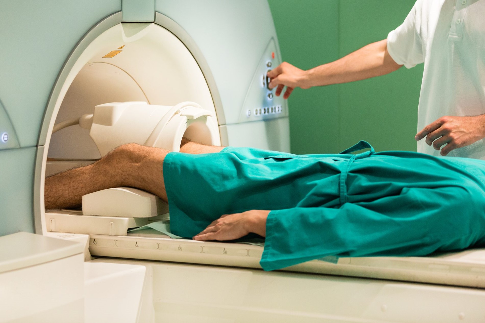
Bone scintigraphy: how it is performed
Bone scintigraphy is a diagnostic technique that allows the graphic representation of the distribution of a radioactive substance, previously injected, in an organ or tissue of the body (the substance is called a tracer)
The scintigraph transforms the emitted radiation into graphic signals
The tracers are injected by intravenous injection (these are phosphate compounds labelled with Tc99m or Iodine-131) and are picked up by the bone matrix in proportion to blood flow and district mineral turnover (e.g. there is a physiological hyperconcentration of tracer at the level of the spine because mineral turnover is higher there).
Bone scintigraphy, when it is performed
The major use of bone scintigraphy is in the search for bone metastases, 50% of which can be detected in the absence of signs on direct radiographic examination.
How to prepare for bone scintigraphy
No preparation is required, you can therefore have breakfast.
However, it is necessary to maintain a high level of hydration throughout the morning, so it seems advisable to have a supply of water.
The time required for the investigation is about 3-4 hours; when the polyphasic method is used, the first two phases immediately follow the administration of the radiopharmaceutical while the late images are acquired after at least 3 hours, after the total-body scan.
It is a good idea to arm yourself with patience and bring something to read.
How bone scintigraphy is performed
The investigation is usually performed by total-body method with a double-headed gammacamera, i.e. with two longitudinal scans of the entire skeleton in a single pass; if necessary, targeted scintigrams are acquired in appropriate projections.
The duration of the total body acquisition is approximately 12 to 15 minutes; that of the individual segments varies between 5 and 15 minutes.
Read Also
Emergency Live Even More…Live: Download The New Free App Of Your Newspaper For IOS And Android
Fusion Prostate Biopsy: How The Examination Is Performed
CT (Computed Axial Tomography): What It Is Used For
What Is An ECG And When To Do An Electrocardiogram
Positron Emission Tomography (PET): What It Is, How It Works And What It Is Used For
Single Photon Emission Computed Tomography (SPECT): What It Is And When To Perform It
Instrumental Examinations: What Is The Colour Doppler Echocardiogram?
Coronarography, What Is This Examination?
CT, MRI And PET Scans: What Are They For?
MRI, Magnetic Resonance Imaging Of The Heart: What Is It And Why Is It Important?
Urethrocistoscopy: What It Is And How Transurethral Cystoscopy Is Performed
What Is Echocolordoppler Of The Supra-Aortic Trunks (Carotids)?
Surgery: Neuronavigation And Monitoring Of Brain Function
Robotic Surgery: Benefits And Risks
Refractive Surgery: What Is It For, How Is It Performed And What To Do?


