Botallo's ductus arteriosus: interventional therapy
Nowadays, there are two techniques for closing the ductus arteriosus of Botallo, both of which are very effective: traditional surgery and transcatheter treatment
The ductus arteriosus is an artery that in foetal life allows blood to flow from the aorta to the pulmonary artery.
When the baby is born, the vessel closes within a few days; if it does not, it is called a pervious ductus arteriosus.
During foetal life, the blood is not oxygenated by the lungs but by the placenta, which also supplies the necessary nutrients to the foetus.
CHILD HEALTH: LEARN MORE ABOUT MEDICHILD BY VISITING THE BOOTH AT EMERGENCY EXPO
During foetal life, the ductus arteriosus of Botallo allows blood to be directed from the pulmonary artery into the aorta
A defining event at birth is the separation of the foetus from the placenta and the reorganisation of the blood circulation.
After the first cry, the lungs expand, becoming capable of exchanging oxygen and carbon dioxide between the environment and the circulation.
The communication created by the duct of Botallo between the aorta and the pulmonary artery will no longer be necessary, and in most newborns this will close within a few days.
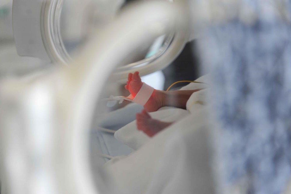
Failure of the Botallo’s duct to close occurs most frequently in premature infants
The share of oxygenated blood passing from the aorta to the pulmonary artery through the ductus arteriosus of Botallo causes an increase in flow and pressure in the ‘left’ part of the heart, which ‘works’ more than normal.
This will result in an increased fluid supply to the lungs causing a condition called pulmonary overflow.
This abnormality is often asymptomatic.
Infants and children with an open (pervious) small ductus arteriosus generally have no symptoms, while symptoms are more often present in preterm infants.
In infants and neonates it can cause fatigability during feeding, growth retardation, recurrent respiratory infections (bronchitis/ bronchopneumonia), and endocarditis (infection of the heart).
Severe cardiac and multi-organ failure may occur in the preterm infant.
If left untreated, the overwork of the heart over decades can lead to:
- Left ventricular dilatation;
- Heart failure;
- Reduced life expectancy.
On auscultation of the heart, the doctor will detect the presence of a continuous heart murmur and will refer the child for an echocardiogram, which is the pivotal examination for the diagnosis of PDA.
QUALITY AED? VISIT THE ZOLL BOOTH AT EMERGENCY EXPO
The chest X-ray and electrocardiogram (ECG) are typically normal.
The electrocardiogram may show left ventricular hypertrophy.
The small Botallo’s duct should ideally be closed in preschool or school age, if large when the diagnosis is made.
Nowadays, there are two techniques for closing the PDA, both of which are very valid: traditional surgery and transcatheter treatment.
The surgical procedure is performed non-open heart and without the need for extracorporeal circulation.
The little patient will require at least one night of intensive care.
Transcatheter treatment is the preferred option when it is feasible, being much less invasive.
It generally does not require an overnight stay in intensive care, and hospitalisation lasts about 3 days.
The procedure is performed via a groin vessel (the femoral artery and/or vein) a small catheter is passed through the aorta to the ductus arteriosus.
After visualisation with contrast medium, the closure device (either a coil or a plug for larger ducts) is brought into place.
Percutaneous treatment can also be performed in premature infants weighing less than 2 kg.
In this particular population of infants, the procedure will be performed using catheters and closure devices of reduced calibre to respect the delicate vascular structures of these patients.
Since a few years there is a new type of device called “Piccolo” used for percutaneous closure of the Botallo’s duct in premature infants.
The dose of contrast medium used will also be reduced to preserve kidney function.
The temperature of the newborns will be kept stable through the use of a heated cot.
Premature neonates may present critical clinical conditions requiring invasive mechanical ventilation, so it will be necessary to transport these patients from the neonatal intensive care unit to the haemodynamic room in an extremely gentle manner.
Regardless of the technique used, closure of the ductus arteriosus without complications equals recovery of the baby.
The day after the transcatheter procedure, the child leaves the hospital and can soon resume all usual activities including sports.
Read Also:
Emergency Live Even More…Live: Download The New Free App Of Your Newspaper For IOS And Android
EMS: Pediatric SVT (Supraventricular Tachycardia) Vs Sinus Tachycardia
Paediatric Toxicological Emergencies: Medical Intervention In Cases Of Paediatric Poisoning
Valvulopathies: Examining Heart Valve Problems
What Is The Difference Between Pacemaker And Subcutaneous Defibrillator?
Heart Disease: What Is Cardiomyopathy?
Inflammations Of The Heart: Myocarditis, Infective Endocarditis And Pericarditis
Heart Murmurs: What It Is And When To Be Concerned
Clinical Review: Acute Respiratory Distress Syndrome
Stress And Distress During Pregnancy: How To Protect Both Mother And Child
Respiratory Distress: What Are The Signs Of Respiratory Distress In Newborns?
What Is Transient Tachypnoea Of The Newborn, Or Neonatal Wet Lung Syndrome?
Tachypnoea: Meaning And Pathologies Associated With Increased Frequency Of Respiratory Acts
Obstructive Sleep Apnoea: What It Is And How To Treat It
Obstructive Sleep Apnoea: Symptoms And Treatment For Obstructive Sleep Apnoea
Transient Tachypnoea Of The Newborn: Overview Of Neonatal Wet Lung Syndrome


