Brain tumours: symptoms, classification, diagnosis and treatment
Brain tumours are neoplastic forms (masses of tissue originating from the uncontrolled proliferation of nerve cells) that can affect the brain, cerebellum or nervous system
The causes of brain tumours are still partly unknown, but a number of risk factors have been identified that increase the likelihood of becoming ill:
- exposure to high doses of radiation;
- exposure to harmful substances (some studies show that these types of tumours occur more frequently in workers in chemical industries or in craftsmen who use solvents);
- infection caused by certain viruses (such as Epstein-Barr).
In addition, some types of brain tumours can run in the same family: in these cases, there is likely to be a hereditary genetic component.
Researchers are investigating other probable causes, such as prolonged use of mobile phones and the presence of post-traumatic brain injury.
Brain tumours can strike at any age, but are most common in children aged 3 to 12 and adults aged 40 to 70.
The manifestations of a brain tumour depend mainly on its location and the size of the tumour mass.
It is caused both by damage to vital tissue and by the pressure exerted by the tumour on the brain.
The most frequent symptoms of a brain tumour are
- headaches (strong in the morning, tending to diminish over the course of the day)
- convulsions;
- hallucinations;
- nausea or vomiting;
- feeling of weakness or reduced sensitivity in the arms or legs;
- lack of coordination when walking;
- abnormal eye movements or visual disturbances (up to blindness);
- drowsiness;
- personality and mood changes;
- memory disorders and confusional states;
- stuttering and speech disorders.
In general, if a neoplasm affects one part of the brain (e.g. the left) the symptom manifests itself in the opposite part (the right): this is due to the fact that each cerebral hemisphere governs the lateral counterpart of the body.
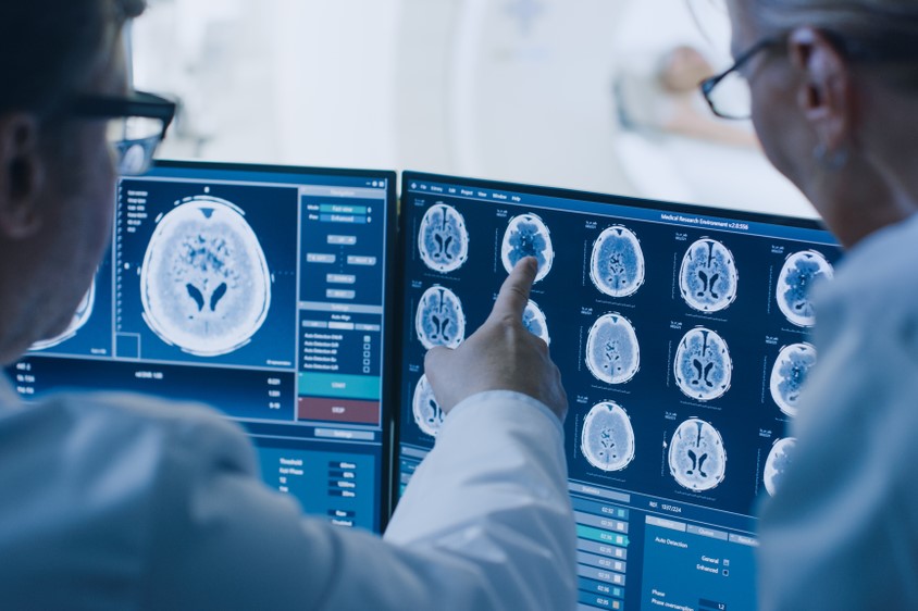
The diagnosis is made by evaluating the patient’s general physical state and a complete neurological examination that assesses cognitive and motor deficits.
Depending on the results, the doctor may request one or more instrumental examinations:
- CT (Computed Axial Tomography);
- MRI (Magnetic Resonance Imaging);
- radiography of the skull;
- electronic brain scan.
Treatment of brain tumours
Given the importance and delicacy of the affected organ and the difficulty of surgery, brain tumours are still among the most difficult to treat.
The treatments used to treat brain tumours are neurosurgery, radiotherapy and chemotherapy.
For benign tumours, neurosurgery can be curative, but for malignant tumours (such as glioblastoma multiforme) the prognosis remains decidedly poor.
In general, neurosurgery serves to reduce the pressure that the tumour exerts inside the skull and thus decrease symptoms; it also enhances the effect of radiotherapy and chemotherapy.
Chemotherapy does not give optimal results, because the brain is very difficult to reach with drugs due to the presence of a natural barrier to external agents.
In recent years, however, research has made progress, adding new chemotherapy drugs and also testing immunotherapy, a technique based on the use of antibodies or cells of the immune system.
Classification: brain tumours can be benign or malignant
Benign tumours are not formed by cancerous cells and have well-defined margins; they are usually removed and in most cases do not recur.
Malignant tumours are formed by cancerous cells, impair vital functions and endanger the survival of the patient. They usually grow very rapidly and invade surrounding tissue.
When a benign brain tumour interferes with vital functions, it is considered malignant even if it is not formed by cancerous cells.
Doctors classify brain tumours according to grade, which can range from low (grade I) to high (grade IV).
The cells of a high-grade tumour present an abnormal appearance and generally grow faster than cells belonging to low-grade tumours.
Brain tumours are also divided into primary (which originate in brain tissue) and secondary (which develop when cancerous cells from another tumour spread to the brain)
In turn, primary tumours are classified according to the tissue in which they originate.
Primary tumours
Gliomas
The most frequent are gliomas, which account for about 40 per cent of all brain tumours.
Gliomas develop from the support cells for the central nervous system (glial cells) that are responsible for performing important functions, such as producing myelin, the white substance that lines nerves and allows nerve impulses to be transmitted.
Medulloblastomas
Malignant tumours among the most frequent in childhood or adolescence.
They originate from primitive (developing) nerve cells that normally disappear from the body after birth.
Most medulloblastomas form in the cerebellum, but can also develop in other areas of the brain.
Meningiomas
They originate in the meninges, i.e. the membranes that surround and protect the brain, and are most common in women between the ages of 30 and 50.
They are generally benign and develop very slowly, allowing the brain to adapt to their presence: this is why meningiomas usually reach a considerable size before they cause symptoms.
Neurinomas
Benign tumours that mainly affect the auditory and trigeminal nerves.
They originate from Schwann cells (hence the name ‘schwannoma’) that cover nerve fibres and are responsible for synthesising myelin (the protective sheath that surrounds nerve cells).
They are tumours of adulthood and affect women more than men.
Hemangioblastomas
They are rare tumours that are usually benign and slow-growing.
They originate in blood vessel cells and are characterised by a familial incidence.
Craniopharyngiomas
Benign tumours that develop in the region of the pituitary gland, located near the hypothalamus, and are most common in children and adolescents.
Generally benign in character, they are sometimes considered malignant as they can compress and damage the hypothalamus impairing vital functions.
Germinomas
Tumours that originate from primitive sex cells or germ cells and spread through the CSF pathways.
They are typical of adolescence and the male sex and respond very well to radiotherapy.
Primary lymphomas
Lymphocytic (white blood cell) derived, they are particularly malignant and common in immunocompromised individuals (such as AIDS patients).
Read Also:
Emergency Live Even More…Live: Download The New Free App Of Your Newspaper For IOS And Android
Thyroid Cancers: Types, Symptoms, Diagnosis
Pediatric Brain Tumors: Types, Causes, Diagnosis And Treatment
Brain Tumours: CAR-T Offers New Hope For Treating Inoperable Gliomas
Lymphoma: 10 Alarm Bells Not To Be Underestimated
Non-Hodgkin’s Lymphoma: Symptoms, Diagnosis And Treatment Of A Heterogeneous Group Of Tumours
CAR-T: An Innovative Therapy For Lymphomas
What Is CAR-T And How Does CAR-T Work?
Symptoms And Treatment For Hypothyroidism
Hyperthyroidism: Symptoms And Causes
Surgical Management Of The Failed Airway: A Guide To Precutaneous Cricothyrotomy
Thyroid Cancers: Types, Symptoms, Diagnosis
Raising The Bar For Pediatric Trauma Care: Analysis And Solutions In The US


