Broken rib (rib fracture): symptoms, causes, diagnosis and treatment
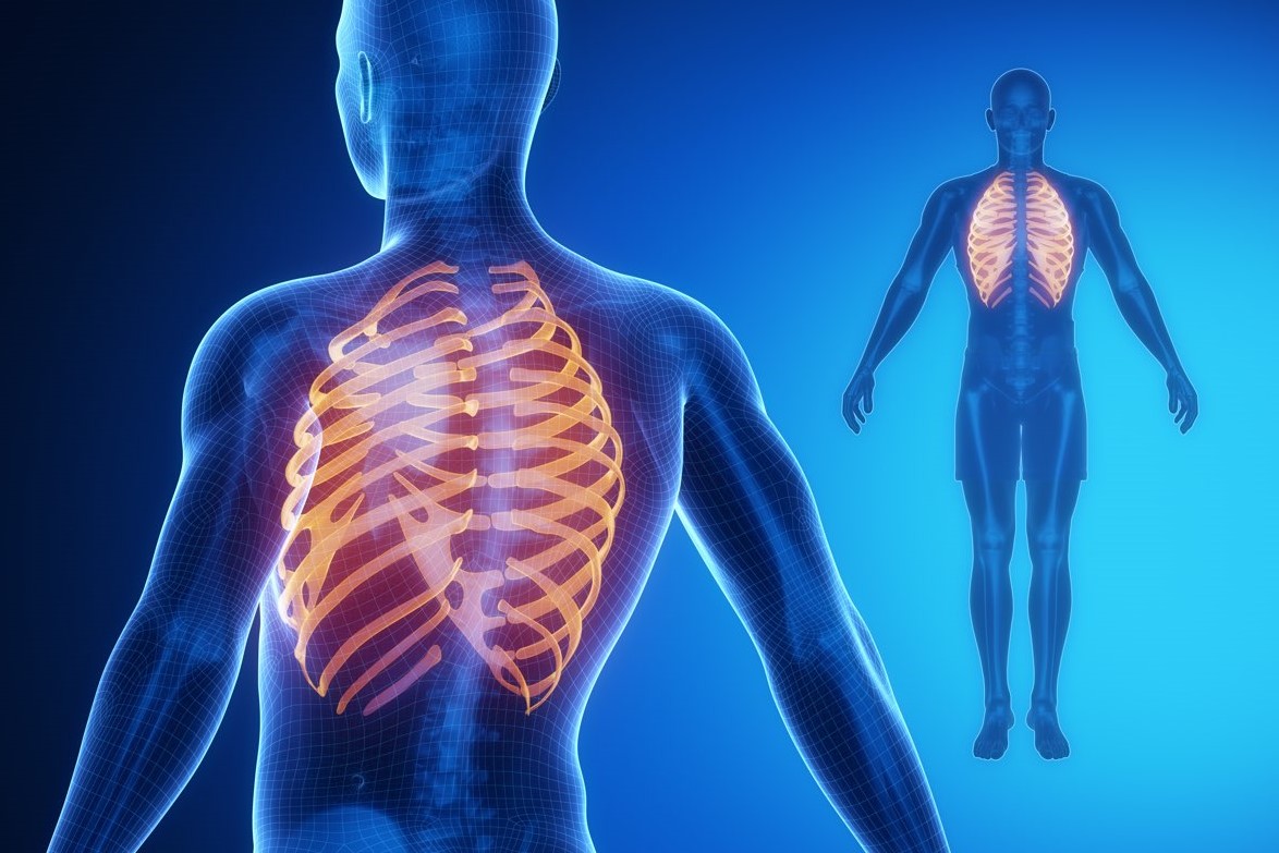
A rib fracture is a fairly common injury, consisting of the more or less serious fracture of the ribs of the chest
Often the fracture affects only one rib; however, in particularly unfortunate cases, it can affect several adjacent ribs simultaneously (multiple rib fracture)
The ribs that most often suffer a fracture are those located in the centre of the rib cage. Fractures of the upper ribs (first and second) usually follow facial trauma or blows to the head.
Causes of a rib fracture
The most common cause of a rib fracture is severe trauma to the chest.
Trauma of such intensity as to break one or more ribs may occur during a car accident, a fall or a playground collision while practising a sport.
In addition to traumatic events, rib fractures can also occur:
- A very loud cough. It may sound strange, but particularly violent coughs can lead to a fracture of the bones that make up the rib cage.
- A repetitive movement at work or during a sport. At these junctures, doctors more appropriately speak of a stress rib fracture. Two possible sports activities that can induce a stress rib fracture are golf and rowing.
Risk factors for a rib fracture
Risk factors for a rib fracture include:
- Osteoporosis. Osteoporosis is a systemic skeletal disease, which causes severe weakening of the bones. This weakening results from a reduction in bone mass, which, in turn, is a consequence of the deterioration of the microarchitecture of bone tissue. Therefore, people with osteoporosis are more prone to fractures because they have more fragile bones than normal.
- Participation in contact sports. Playing sports in which physical contact is involved carries a high risk of fractures, not only in the lower or upper limbs, but also in the chest. The sportsmen and women most at risk are rugby, football, American football, ice hockey and basketball players.
- Neoplastic lesions of the ribs. A malignant tumour originating in a rib weakens the rib, making it more fragile and particularly susceptible to fractures.
Symptoms and complications
The characteristic symptom of a fracture is localised pain at the point of bone breakage.
The pain sensation varies from patient to patient, depending on location, number of ribs affected and individual pain tolerance.
Post-fracture pain in the ribs tends to worsen in some particular circumstances:
- When the patient is breathing deeply.
- With compression of the injured chest area.
- With twisting and bending movements of the body.
If, due to very intense pain, the patient cannot breathe normally, he/she tends to suffer from:
- Shortness of breath
- Headaches
- Dizziness, light-headedness, tiredness and/or drowsiness
- Anxiety and restlessness
Often, when the cause of the fracture is trauma, two signs appear on the thoracic area involved in the impact that certainly do not go unnoticed: swelling and haematoma.
Multiple fracture: what are the risks?
If the rib fracture is multiple, it can lead to a potentially fatal medical condition, identified by the term ‘rib volet’.
When to seek medical attention?
If they experience severe and permanent pain and have trouble breathing, people who have suffered severe chest trauma should seek medical attention or go to the nearest hospital.
Complications in a rib fracture
If severe or untreated, a fracture of one or more ribs can lead to several complications, including:
- Injury of the major thoracic blood vessels. This occurs when the rupture affects the first three pairs of upper ribs. Damage to the aorta or the other major vessels of the thorax is caused by one of the two pointed bone stumps resulting from the fracture.
- Injury to one of the lungs. The ribs that, if fractured, can damage the lungs are those located in the middle of the rib cage. As before, it is one of the two sharp bony stumps, which are created after a broken bone, that ‘stings’ the lungs. The main consequence of a rib injuring a lung is the collapse of the lung itself, due to air and blood entering the pleural cavity. In medicine, this condition is also known as pneumothorax (PNX).
- Injury to the spleen, liver or kidneys. These three organs are at risk of damage when the fracture affects the lower ribs and is such that it creates very sharp extremities.
- Pneumonia and other pulmonary disorders. The inability to breathe deeply, because this causes pain, can lead to the onset of even severe lung inflammation.
Differences from cracked rib
The symptomatic aspect that most differentiates a rib fracture from a crack is the fact that, in the latter case, there is no risk of injury to the internal organs of the chest.
Diagnosis
Generally, the diagnostic procedure for detecting a rib fracture involves, firstly, a thorough objective examination and, secondly, the performance of a series of instrumental examinations, in some cases quite invasive.
Since a fractured rib can lead to some dangerous complications, diagnosing it correctly is very important.
This explains why doctors, in the presence of rib pain, are particularly scrupulous in wanting to understand the exact cause of the present symptomatological picture.
OBJECTIVE EXAMINATION
During the objective examination, the doctor examines the patient, looking for any external clinical signs (haematomas, swelling, etc.), and questions him/her about the symptoms:
- What are they?
- Following what event did they appear?
- What movements or gestures exacerbate their intensity?
Questions of this kind make it possible to understand, in broad terms, the underlying problem and what caused it.
After the questionnaire, the objective examination ends with palpation of the painful area (to see what the patient’s response is), auscultation of the lungs and heart (looking for any abnormal sounds), and examination of the head, neck, spinal cord and abdomen.
INSTRUMENTAL EXAMINATIONS
Instrumental examinations are essential, as the information they provide allows a correct and safe final diagnosis to be reached.
Prescribed procedures may include:
- X-rays. They allow the detection of most rib fractures. In fact, they have limitations only in the presence of ‘fresh’ and not clean rib fractures. X-rays are ionising radiation that is harmful to health; however, it should be remembered that the dose of such radiation is minimal.
- CT SCAN. It provides a series of three-dimensional images that reproduce the internal anatomy of the body very clearly. It is very useful for analysing not only the bones of the entire rib cage, but also the health of the thoracic blood vessels, lungs and abdominal organs. It relies on the use of non-negligible amounts of ionising radiation.
- Nuclear magnetic resonance imaging (NMR). This is a radiological examination that involves exposing the patient to completely harmless magnetic fields, without the need for harmful ionising radiation. Like CT, it is useful for evaluating a wide range of elements: ribs, blood vessels passing through the chest, lungs and organs of the abdomen.
- Bone scintigraphy. This is a very sensitive nuclear medicine examination, as it shows any bone changes, even the least obvious. Precisely because of its sensitivity, doctors prescribe it when they suspect minimal fractures, which are not visible with previous instrumental examinations. Such fractures are those that a repetitive gesture or a loud cough can cause. Unfortunately, it is a rather invasive diagnostic technique. In fact, it involves the venous injection of a radioactive drug.
Treatment of rib fracture
The treatment that doctors adopt in the case of a rib fracture involves rest, applying ice to the painful area, and taking pain-relieving drugs.
Among the most prescribed painkillers are aspirin, aspirin derivatives and ibuprofen.
Read Also:
Emergency Live Even More…Live: Download The New Free App Of Your Newspaper For IOS And Android
Facial Trauma With Skull Fractures: Difference Between LeFort Fracture I, II And III
Airway Management After A Road Accident: An Overview
Tracheal Intubation: When, How And Why To Create An Artificial Airway For The Patient
What Is Transient Tachypnoea Of The Newborn, Or Neonatal Wet Lung Syndrome?
Traumatic Pneumothorax: Symptoms, Diagnosis And Treatment
Diagnosis Of Tension Pneumothorax In The Field: Suction Or Blowing?
Pneumothorax And Pneumomediastinum: Rescuing The Patient With Pulmonary Barotrauma
Difference Between Compound, Dislocated, Exposed And Pathological Fracture
Cervical Collar In Trauma Patients In Emergency Medicine: When To Use It, Why It Is Important
KED Extrication Device For Trauma Extraction: What It Is And How To Use It
Primary, Secondary And Hypertensive Spontaneous Pneumothorax: Causes, Symptoms, Treatment
Penetrating And Non-Penetrating Cardiac Trauma: An Overview


