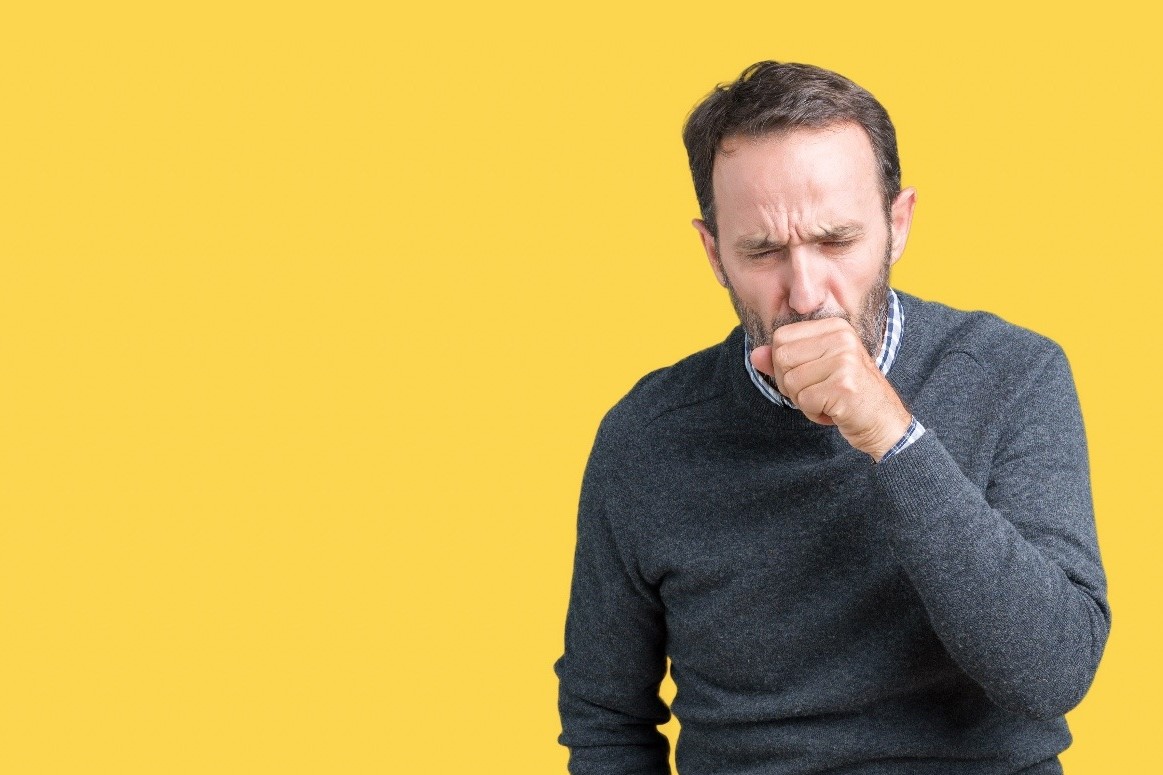
Bronchospasm: symptoms, causes and treatment
Bronchospasm refers to an unusual and excessive contraction of the smooth muscles that surround the bronchi and bronchioles
This condition results in narrowing, which can lead to airway obstruction.
These are usually temporary problems; later there is a restoration of airway patency.
Unfortunately, sufferers of this discomfort cannot breathe well, cough often, and have wheezes during breathing.
He or she may also complain of chest tightness and oppression.
The large mucus production by the mucous membrane of occluded bronchi and bronchioles determine coughing as an annoying cause of bronchospasm.
The main causes of bronchospasm are asthma and bronchitis, inflammatory conditions
Generally, the therapy proposed to relieve symptoms from bronchoplasmos is pharmacological and consists of airway-opening medications.
For example, beta2-agonists and anticholinergic bronchodilators or anti-inflammatory drugs for reducing the inflammatory state, such as corticosteroids, may be prescribed.
What are the symptoms of bronchospasm?
The symptoms that indicate the presence of bronchospasm are definitely the following:
- coughing due to excessive accumulation of mucus produced by the mucosa of the bronchi and bronchioles;
- shortness of breath and dyspnea. These breathing-related difficulties worsen in the evening, in the morning or after physical activity for those who already suffer from asthma or chronic bronchitis.
- sensation of chest occlusion, which can cause real chest pain and tightness;
- rales during breathing.
Bronchospasm in children
Obviously, younger children are more susceptible to developing bronchospasm because they have a smaller bronchial lumen, in which even modest changes in the airway walls can greatly limit the amount of air able to reach the lungs.
Generally, this easier exposure in children resolves as the child grows.
During childhood, up to age 6, respiratory infections of viral (respiratory syncytial virus) or bacterial (pneumococcal, haemophilus influenzae) type are the main cause.
These agents inflame the bronchial wall with irritation of the smooth muscle, which contracts in response.
Causes
The main factors causing bronchospasm are essentially two well-known inflammatory conditions of the bronchial tree: asthma and bronchitis.
Asthma
Asthma is a chronic morbid condition, most likely of genetic derivation.
Its symptoms are usually amplified after contact with allergens (substances that the body considers foreign and potentially dangerous, therefore deserving of an immune attack in order to be eliminated), such as pollen, particular foods, dust, animal dander, medicines.
Physical exertion, excessive emotions, anxiety, stress, and smoking can also exacerbate asthma.
Bronchitis
Bronchitis unlike asthma, on the other hand, can be an acute or chronic circumstance that emerges due to respiratory infections.
For example, it may be a consequence of a cold or flu, cigarette smoking, and/or environmental, household, or occupational pollution.
Other causes
General anesthesia, practiced in surgery before certain very invasive procedures, can also be a cause of bronchospasm.
In this case, the difficulty related to breathing is a complication of anesthesia.
Its occurrence is in fact subsequent to the physician’s application of the tube used to support the patient’s breathing, during the operation.
Excessive physical activity, at levels too lax compared to the subject’s capabilities, can also lead to bronchospasm.
The main consequences of bronchospasm
What are the consequences and complications suffered by those suffering from bronchospasm?
Definitely fatigue in breathing, as there is an impediment to the passage of air through bronchi and/or bronchioles.
As mentioned above, however, the situation appears more complex because of the excessive mucus that the bronchial mucosa produces, which consequently results in:
- little air enters the lungs;
- irritation of the inner wall of the bronchi or bronchioles, which becomes inflamed;
- coughing, as a defensive action of the body trying to expel the mucus in the bronchi.
Asphyxia
If bronchospasm is neglected and becomes very acute, it can even lead to the death of the individual from asphyxia.
The clinical manifestations that characterize the presence of severe respiratory distress are dyspnea at rest, cyanosis (usually in the fingers) and increased heart rate.
When to seek medical advice?
It is important to seek medical attention when the cough does not go away and the wheezing while the person is breathing worsens.
Also, if there is the presence of a febrile state and problems related to breathing.
In addition, again according to expert opinion, these are symptoms that require immediate medical examination:
- coughing with the presence of blood;
- dyspnea and cyanosis in the fingers;
- severe chest pain;
- increased heartbeats.
Diagnosis
If bronchospasm is suspected, the physician, before testing, resorts to objective test and evaluation of the patient’s medical history.
These two tests are generally sufficient to establish an accurate final diagnosis.
However, more specific instrumental tests may also need to be performed, as these also clarify the underlying causes of the bronchospasm episodes.
Instrumental tests for recognizing bronchospasm
The instrumental tests mentioned above that the physician may subject the patient to in order to understand the underlying causes of the discomfort are:
- Rx-chest. This test is used to provide a fairly sharp image of the state of the lungs and other structures inside the chest. In this way, the presence of a possible lung infection can be checked.
- CT scan – Computed Axial Tomography. This is used to give the specialist very comprehensive three-dimensional images of the organs contained in the chest cavity. In this way all abnormalities, which may affect the lungs, can be diagnosed. For example, signs of infection, signs of inflammation, etc. However, the patient with CT scan is exposed to ionizing radiation, so it is considered an invasive test not suitable for children and pregnant women. In some cases, to increase image quality, the doctor administers a contrast medium into the patient’s circulatory stream. If used, this substance increases the level of invasiveness of the test. Allergic reactions could be triggered.
- Finally, spirometry, which is very quick and rapid compared to the first two, is used to record the inspiratory and expiratory capacity of the lungs, as well as the opening of the airways passing through them.
Read Also
Emergency Live Even More…Live: Download The New Free App Of Your Newspaper For IOS And Android
Ventilator Management: Ventilating The Patient
Three Everyday Practices To Keep Your Ventilator Patients Safe
Ambulance: What Is An Emergency Aspirator And When Should It Be Used?
The Purpose Of Suctioning Patients During Sedation
Supplemental Oxygen: Cylinders And Ventilation Supports In The USA
Basic Airway Assessment: An Overview
Respiratory Distress: What Are The Signs Of Respiratory Distress In Newborns?
EDU: Directional Tip Suction Catheter
Suction Unit For Emergency Care, The Solution In A Nutshell: Spencer JET
Airway Management After A Road Accident: An Overview
Tracheal Intubation: When, How And Why To Create An Artificial Airway For The Patient
What Is Transient Tachypnoea Of The Newborn, Or Neonatal Wet Lung Syndrome?
Traumatic Pneumothorax: Symptoms, Diagnosis And Treatment
Diagnosis Of Tension Pneumothorax In The Field: Suction Or Blowing?
Pneumothorax And Pneumomediastinum: Rescuing The Patient With Pulmonary Barotrauma
ABC, ABCD And ABCDE Rule In Emergency Medicine: What The Rescuer Must Do
Multiple Rib Fracture, Flail Chest (Rib Volet) And Pneumothorax: An Overview
Internal Haemorrhage: Definition, Causes, Symptoms, Diagnosis, Severity, Treatment
Assessment Of Ventilation, Respiration, And Oxygenation (Breathing)
Oxygen-Ozone Therapy: For Which Pathologies Is It Indicated?
Difference Between Mechanical Ventilation And Oxygen Therapy
Hyperbaric Oxygen In The Wound Healing Process
Venous Thrombosis: From Symptoms To New Drugs
What Is Intravenous Cannulation (IV)? The 15 Steps Of The Procedure
Nasal Cannula For Oxygen Therapy: What It Is, How It Is Made, When To Use It
Nasal Probe For Oxygen Therapy: What It Is, How It Is Made, When To Use It
Oxygen Reducer: Principle Of Operation, Application
How To Choose Medical Suction Device?
Holter Monitor: How Does It Work And When Is It Needed?
What Is Patient Pressure Management? An Overview
Head Up Tilt Test, How The Test That Investigates The Causes Of Vagal Syncope Works
Cardiac Syncope: What It Is, How It Is Diagnosed And Who It Affects
Cardiac Holter, The Characteristics Of The 24-Hour Electrocardiogram


