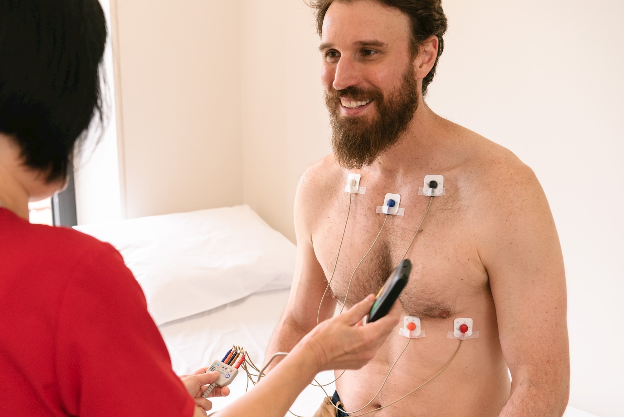
Cardiac Holter, the characteristics of the 24-hour electrocardiogram
What is the cardiac Holter? An electrocardiogram is a diagnostic test designed to record cardiac activity in order to assess the health of the heart and detect abnormalities, alterations in heart rhythm or heart disease of various kinds
A special instrument called an electrocardiograph is used to perform an ECG, which is able to monitor cardiac function and report it graphically in the form of a tracing.
Depending on the patient’s needs, the cardiologist may prescribe different types of ECGs:
- the standard ECG is used to measure cardiac activity under normal conditions; it involves placing 12 to 15 electrodes on the patient’s chest, arms and legs. The recording lasts a few minutes during which it is sufficient to remain lying down while breathing regularly and avoiding movement or talking;
- the exercise ECG assesses changes in cardiac activity when the heart is subjected to physical activity; after electrodes are applied, the measurement usually involves performing some simple exercises, such as pedaling on an exercise bike or running on a treadmill. The duration of the recording can range from 10 to 40 minutes depending on the specific case; alternatively, in some cases, special drugs can be administered to simulate the effects of physical activity. The exercise ECG can, in addition, be used to monitor the effects of particular drug therapies on the heart;
- the dynamic ECG according to Holter allows monitoring of cardiac function over a specific period of time that generally lasts 24 to 48 hours. Cardiac Holter is performed using a special hand-held device, which is connected to a series of electrodes placed on the patient’s chest and allows the electrical activity of the heart to be recorded, and then the tracing is extrapolated at a later time.
Why is a dynamic electrocardiogram (cardiac Holter) performed?
The 24-hour ECG is usually prescribed to diagnose heart rhythm changes characterized by sporadic and discontinuous occurrence, which could not be missed by a standard ECG.
In some cases, it also enables the evaluation of the effectiveness of drug therapies undertaken to treat certain cardiac diseases and disorders, or to monitor the functioning of implanting devices such as pacemakers, cardioconverters, or defibrillators.
How should I prepare for cardiac holter?
In general, the electrocardiogram is a noninvasive test that requires no special preparation.
On the day of the procedure, however, the physician may communicate a number of useful instructions to the patient:
- first, care should be taken not to accidentally remove the electrodes for the duration of the examination; to this end, it is advisable to avoid particular sports activities and refrain from taking showers or baths;
- in general, it is important to conduct a typical day normally, performing all the activities that one usually does. Any variations in one’s habits, in fact, could lead to false and inaccurate results;
- another useful expedient may be to keep a diary in which to note any moments of the day that may have induced palpitations, chest pain, dizziness or breathlessness. In this way, the cardiologist will be able to determine more accurately what are the underlying causes of any conditions.
*This is approximate information; therefore, it is necessary to contact the facility where the examination is being performed to obtain specific information on the preparation procedure.
Read Also
Emergency Live Even More…Live: Download The New Free App Of Your Newspaper For IOS And Android
Holter Monitor: How Does It Work And When Is It Needed?
What Is Patient Pressure Management? An Overview
Head Up Tilt Test, How The Test That Investigates The Causes Of Vagal Syncope Works
Cardiac Syncope: What It Is, How It Is Diagnosed And Who It Affects
Holter Blood Pressure: What Is The ABPM (Ambulatory Blood Pressure Monitoring) For?
Head Up Tilt Test, How The Test That Investigates The Causes Of Vagal Syncope Works
Aslanger Pattern: Another OMI?
Abdominal Aortic Aneurysm: Epidemiology And Diagnosis
What Is The Difference Between Pacemaker And Subcutaneous Defibrillator?
Heart Disease: What Is Cardiomyopathy?
Inflammations Of The Heart: Myocarditis, Infective Endocarditis And Pericarditis
Heart Murmurs: What It Is And When To Be Concerned
Clinical Review: Acute Respiratory Distress Syndrome
Botallo’s Ductus Arteriosus: Interventional Therapy
Heart Valve Diseases: An Overview
Cardiomyopathies: Types, Diagnosis And Treatment
First Aid And Emergency Interventions: Syncope
Tilt Test: What Does This Test Consist Of?
Cardiac Syncope: What It Is, How It Is Diagnosed And Who It Affects
New Epilepsy Warning Device Could Save Thousands Of Lives
Understanding Seizures And Epilepsy
First Aid And Epilepsy: How To Recognise A Seizure And Help A Patient
Neurology, Difference Between Epilepsy And Syncope
Positive And Negative Lasègue Sign In Semeiotics
Wasserman’s Sign (Inverse Lasègue) Positive In Semeiotics
Positive And Negative Kernig’s Sign: Semeiotics In Meningitis
Lithotomy Position: What It Is, When It Is Used And What Advantages It Brings To Patient Care
Trendelenburg (Anti-Shock) Position: What It Is And When It Is Recommended
Prone, Supine, Lateral Decubitus: Meaning, Position And Injuries
Stretchers In The UK: Which Are The Most Used?
Does The Recovery Position In First Aid Actually Work?
Reverse Trendelenburg Position: What It Is And When It Is Recommended
Drug Therapy For Typical Arrhythmias In Emergency Patients
Canadian Syncope Risk Score – In Case Of Syncope, Patients Are Really In Danger Or Not?
What Is Ischaemic Heart Disease And Possible Treatments
Percutaneous Transluminal Coronary Angioplasty (PTCA): What Is It?
Ischaemic Heart Disease: What Is It?
EMS: Pediatric SVT (Supraventricular Tachycardia) Vs Sinus Tachycardia
Paediatric Toxicological Emergencies: Medical Intervention In Cases Of Paediatric Poisoning
Valvulopathies: Examining Heart Valve Problems


