Cardiac Rhythm Disturbance Emergencies: the experience of US rescuers
Learn how US EMTs and paramedics identify, treat and care for patients with cardiac rhythm disorders
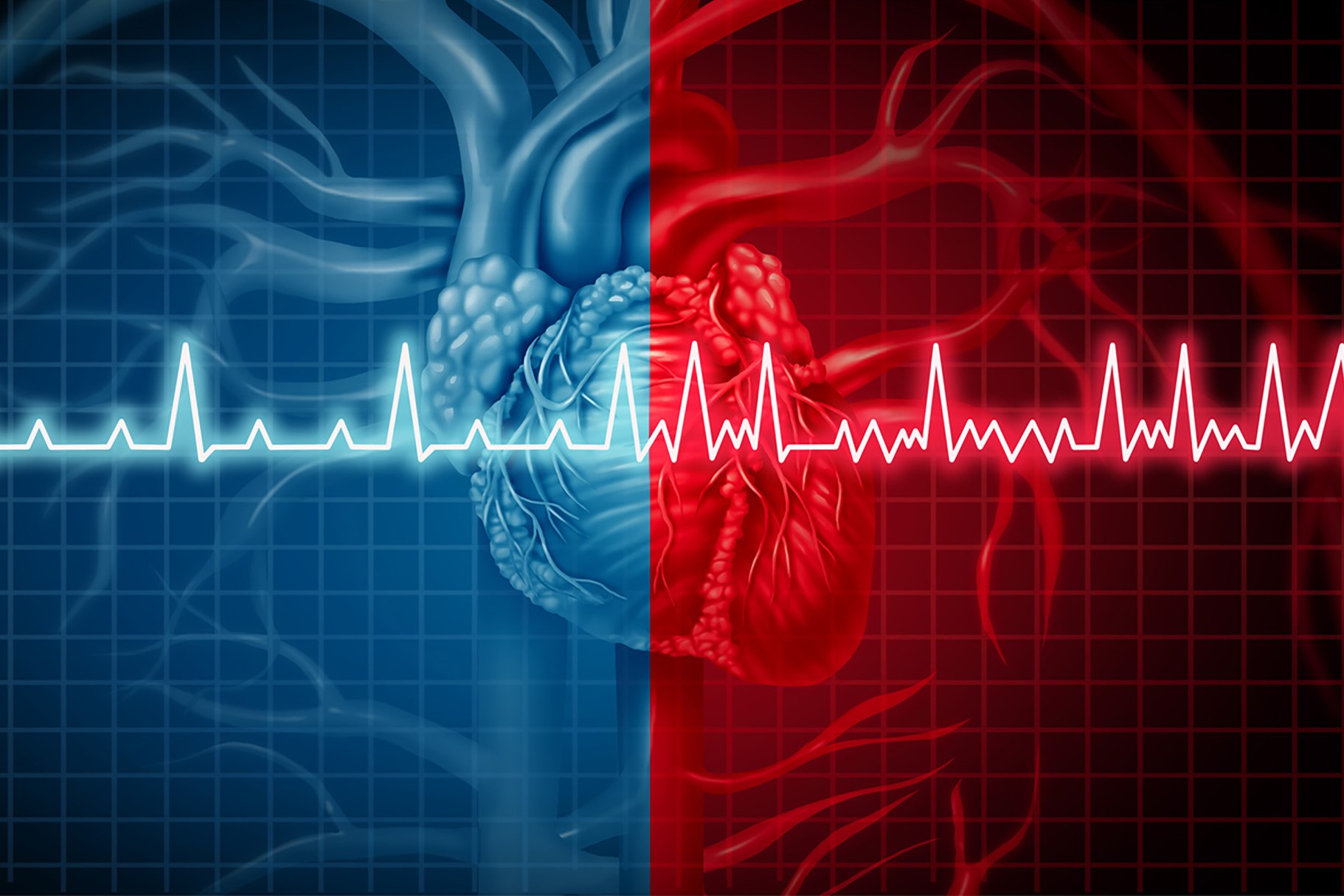
A cardiac rhythm disturbance, also known as arrhythmia, occurs when there is a problem with the rate or rhythm of the heartbeat
During an arrhythmia, the heart beats too fast, too slow, or irregularly.
When a heart beats too fast, it’s called tachycardia, and when a heart beats too slow, it’s called bradycardia.
Arrhythmia is caused by changes in heart tissue or in the electrical signals that control the heartbeat.
These changes may occur because of disease, injury, or genetics.
QUALITY AED? VISIT THE ZOLL BOOTH AT EMERGENCY EXPO
In many cases, patients with arrhythmia do not experience any symptoms.
However, in other cases, you may feel an irregular heartbeat, faint, dizzy, or have difficulty breathing.
Arrhythmia affects millions of people and is associated with substantial morbidity. Atrial fibrillation, or AFib, is one of the most common arrhythmias, and it affects about 2.3 million people in the United States today.
About cardiac rhythm disorders: What is Arrhythmia?
Cardiac rhythm disturbance, also known as arrhythmia, is a group of conditions in which the heartbeat is irregular, too fast, or too slow.
When a patient has a heart rate of more than 100 beats per minute, this is called tachycardia.
When a patient has a heart rate of fewer than 60 beats per minute, this is called bradycardia.
Symptoms of arrhythmia, when present, may include palpitations or feeling a pause between heartbeats.
There may be lightheadedness, passing out, shortness of breath, or chest pain in more severe cases.
STRETCHERS, LUNG VENTILATORS, EVACUATION CHAIRS: SPENCER PRODUCTS ON DOUBLE BOOTH AT EMERGENCY EXPO
While most types of arrhythmia are not serious, some forms predispose a person to complications such as stroke or heart failure.
Others may even result in sudden death.
Cardiac Rhythm Disorders, Where Are Four Main Types of Arrhythmia:
- Bradyarrhythmia, also called bradycardia, is a slow heart rate. For adults, bradycardia is often defined as a heart rate that is slower than 60 beats per minute, although some studies use a heart rate of fewer than 50 beats per minute. Some people, especially people who are young or physically fit, may generally have slow heart rates. Visit your doctor in order to determine whether a slow heart rate is appropriate for you.
- Premature or extra heartbeat is the most common form of arrhythmia. This type of arrhythmia occurs when the signal for the heart to beat comes early. It can feel like your heart fluttered or skipped a beat. The premature or extra heartbeat creates a short pause, followed by a stronger beat when your heart returns to its regular rhythm. There are often no symptoms of a premature or extra heartbeat. Premature heart rhythm most frequently happens naturally but can be associated with caffeine and nicotine consumption or stress. Usually, no treatment is required.
- Supraventricular arrhythmia. These forms of arrhythmia start in the heart’s upper chambers, called the atrium, or at the gateway to the lower chambers. Supraventricular arrhythmias result in fast heart rates or tachycardia. Tachycardia occurs when the heart, at rest, goes over 100 beats per minute. This fast pace is sometimes paired with an uneven heart rhythm.
There are several different forms of supraventricular arrhythmias, including:
A) Atrial fibrillation (AFib). This is one of the most common types of arrhythmia. The heart can race at more than 400 beats per minute.
B) Atrial flutter. Atrial flutter can cause the upper chambers to beat 250 to 350 times per minute. The signal that tells the upper chambers to beat may be disrupted when it encounters damaged tissue, such as a scar. The signal may find an alternate path, creating a loop that causes the upper chamber to beat repeatedly. As with atrial fibrillation, some but not all of these signals travel to the lower chambers. As a result, the upper chambers and lower chambers beat at different rates.
C) Paroxysmal supraventricular tachycardia (PSVT). In PSVT, electrical signals that begin in the upper chambers and travel to the lower chambers cause extra heartbeats. The arrhythmia begins and ends suddenly. It can happen during vigorous physical activity. It is usually not dangerous and tends to occur in young people.
- Ventricular arrhythmia. These arrhythmias start in the heart’s lower chambers. They can be very dangerous and usually require medical care right away.
A) Ventricular tachycardia is a fast, regular beating of the ventricles that may last for only a few seconds or much longer. A few beats of ventricular tachycardia often do not cause problems. However, episodes that last for more than a few seconds can be dangerous. Ventricular tachycardia can turn into more serious arrhythmias, such as ventricular fibrillation or v-fib.
B) Ventricular fibrillation (V-fib) occurs when disorganized electrical signals make the ventricles quiver instead of pumping normally. Without the ventricles pumping blood to the body, sudden cardiac arrest and death can occur within a few minutes.
C) Torsades de pointes is a type of ventricular fibrillation that develops in people with Long QT syndrome, a rare disorder of the heart’s electrical system. Torsades de pointes causes a rapid heartbeat, which restricts oxygen-rich blood flow. The lack of oxygen can cause sudden fainting. Short episodes of torsades de pointes (less than 1 minute) often resolve themselves so that patient regains consciousness. If the episode lasts longer, it can lead to VFib and serious complications.

How Are Arrhythmias Diagnosed?
If you have symptoms of arrhythmia, you should make an appointment with a cardiologist immediately.
After evaluating your symptoms, the cardiologist may perform a variety of diagnostic tests, including:
- Electrocardiogram (ECG or EKG):A picture of the electrical impulses traveling through the heart muscle.
- Stress test: A test used to record arrhythmias that start or are worsened with exercise.
- Echocardiogram: A type of ultrasound that provides a view of the heart to determine if there is heart muscle or valve disease.
- Cardiac catheterization: Using a local anesthetic, a catheter (small, hollow, flexible tube) is inserted into a blood vessel and guided to the heart so X-ray videos of your arteries, heart chambers, and valves can be taken to show how well your heart muscle and valves are working.
- Electrophysiology study (EPS): A special heart catheterization that evaluates your heart’s electrical system.
- Tilt table test: This test records your blood pressure and heart rate on a minute-by-minute basis while you lay on a table that is tilted to evaluate your condition as you change positions.
Risk Factors for Cardiac Rhythm Disorders (Arrhythmia)
The following factors may cause you to have an increased risk for arrhythmia:
- Age: The chances of having arrhythmia grow as we age, in part because of changes in heart tissue and in how the heart works overtime. Older people are also more likely to have health conditions, including heart disease, that raise the risk of arrhythmia.
- Environment: Exposure to air pollutants, especially particulates and gases, is linked to short-term risk of arrhythmia.
- Family history and genetics: If your parent or other close relative has had arrhythmia, you may have an increased risk for arrhythmia. Also, some inherited types of heart disease can increase your risk of arrhythmia.
- Lifestyle habits: Your risk for arrhythmia may be higher because of certain lifestyle habits, including: Drinking alcohol, Smoking, Using illegal drugs, such as cocaine or amphetamines
- Race or ethnicity: Studies suggest that Caucasian Americans may be more likely to have some forms of arrhythmia than African Americans, such as atrial fibrillation.
- Sex: Studies suggest that men are more likely to have atrial fibrillation than women. However, women taking certain medicines appear to be at a higher risk of certain types of arrhythmia.
- Surgery: You may be at higher risk of developing atrial flutter in the early days and weeks after surgery involving the heart, lungs, or esophagus.
- Other medical conditions: Arrhythmias are more common in people who have diseases or conditions that weaken the heart, but many conditions can raise the risk for arrhythmia. These include:
A) Aneurysms
B) Autoimmunedisorders, such as rheumatoid arthritis and lupus
C) Cardiomyopathy
D) Diabetes
F) Eating disorders, such as bulimia and anorexia
G) Heart attack
H) Heart inflammation
I) Heart failure
L) Heart tissue that is too thick or stiff or that has not formed normally.
M) High blood pressure
N) Viral infections such as influenza or COVID-19.
O) Kidney disease
P) Leaking or narrowed heart valves
Q) Low blood sugar
R) Lung diseases
S) Musculoskeletal disorders
T) Obesity
U) Overactive or underactive thyroid gland
V) Sepsis
Z) Sleep apnea
How to Prevent Arrhythmia
Many arrhythmias are not a serious condition and can be prevented by avoiding known triggers, including:
- Certain emotions, such as anxiety, stress, panic, and fear
- Too much caffeine
- Nicotine from cigarettes or e-cigarettes
- Illegal drugs, such as cocaine
- Diet pills
- Increased exercise
- A fever
Signs and Symptoms Arrhythmia (cardiac rhythm disorders)
An arrhythmia may not cause any noticeable symptoms.
If symptoms occur, they may include:
- Palpitations: A feeling of skipped heartbeats, fluttering, “flip-flops,” or feeling that the heart is “running away.”
- Pounding in the chest
- Dizziness or feeling lightheaded
- Shortness of breath
- Chest discomfort
- Weakness or fatigue (feeling very tired)
If you have symptoms of arrhythmia, keep track of when and how often they occur to help your doctor diagnose and treat your condition.
If left untreated, arrhythmia can lead to life-threatening complications such as stroke, heart failure, or sudden cardiac arrest.
When to Call Emergency Number for Arrhythmia
If you experience an abnormal heartbeat that does not go back to normal within a few minutes, or your symptoms worsen, call your doctor.
Call Emergency Number right away if you have any of these symptoms:
- Pain or pressure in the middle of your chest that lasts more than a few minutes
- Pain that spreads to your jaw, neck, arms, back, or stomach
- Nausea
- Cold sweat
- Drooping face
- Arm weakness
- Trouble speaking
How are Arrhythmias Treated?
Most arrhythmias are treatable. Treatments may include the following:
Healthy lifestyle changes: Your doctor may recommend that you adopt the following lifelong heart-healthy lifestyle changes to lower your risk for conditions such as high blood pressure and heart disease, which can lead to arrhythmia.
- Aiming for a healthy weight
- Being physically active
- Heart-healthy eating
- Managing stress
- Quitting smoking
Medicines: Your doctor may give you medicine for your arrhythmia.
This might include:
- Adenosine to slow a racing heart.
- Atropine to treat a slow heart rate.
- Beta-blockers to treat high blood pressure or a fast heart rate or to prevent repeat episodes of arrhythmia.
- Blood thinners to reduce the risk of blood clots forming.
- Calcium channel blockers slow a rapid heart rate or the speed at which signals travel.
- Digitalis, or digoxin, to treat a fast heart rate.
- Potassium channel blockers slow the heart rate.
- Sodium channel blockers block electrical signal transmission, lengthen cell recovery periods, and make cells less excitable.
Procedures: If medicines do not treat your arrhythmia, your doctor may recommend one of these procedures or devices.
- Cardioversion
- Catheter ablation
- Implantable cardioverter defibrillators (ICDs)
- Pacemakers
Other treatments: Treatment may also include managing any underlying condition, such as an electrolyte imbalance, high blood pressure, heart disease, sleep apnea, or thyroid disease.
How Do EMTs & Paramedics Treat Arrhythmia?
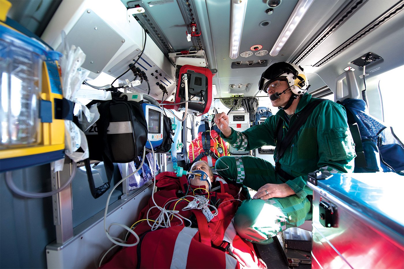
In the event of an arrhythmia emergency, an EMT or paramedic will likely be the first healthcare provider to assess and treat your condition.
EMTs have a clear set of protocols and procedures for most of the 911 emergencies they encounter, including arrhythmia.
For all suspected arrhythmias, the first step is a rapid and systematic assessment of the patient.
For this assessment, most EMS providers will use the ABCDE approach.
ABCDE stands for Airway, Breathing, Circulation, Disability, and Exposure.
The ABCDE approach is applicable in all clinical emergencies for immediate assessment and treatment.
It can be used in the street with or without any equipment.
It can also be used in a more advanced form where emergency medical services are available, including emergency rooms, hospitals, or intensive care units.
Cardiac Rhythm Disorders: Treatment Guidelines & Resources for Medical First Responders
The National Model EMS Clinical Guidelines by the National Association of State EMT Officials (NASEMSO) provides treatment guidelines for bradycardia on page 30 and tachycardia with a pulse on page 37.
These guidelines are maintained by NASEMSO to facilitate the creation of state and local EMS system clinical guidelines, protocols, and operating procedures.
These guidelines are either evidence-based or consensus-based and have been formatted for use by EMS professionals.
The guidelines include the following inclusion criteria for bradycardia:
1) Heart rate less than 60 beats per minute with either symptoms (AMS, CP, CHF, seizure, syncope, shock, pallor, diaphoresis) or evidence of hemodynamic instability
2) The major EKG rhythms classified as bradycardia include:
- Sinus bradycardia
- Second-degree AV block
Type I — Wenckebach/Mobitz I
Type II — Mobitz II
- Third-degree AV block complete block
- Ventricular escape rhythms
3) See additional inclusion criteria below for pediatric patients
The guidelines include the following inclusion criteria for tachycardia with a pulse:
Heart rate greater than 100 bpm in adults or relative tachycardia in pediatric patients.
EMS Protocol for Atrial Fibrillation
The American College of Cardiology, the American Heart Association, and the Heart Rhythm Society released the “2019 AHA/ACC/HRS Focused Update of the 2014 Guideline for Management of Patients with Atrial Fibrillation.”
The purpose of these guidelines is to update the 2014 AF Guideline where new evidence has emerged. A free copy of this clinical practice guideline can be downloaded here.
National-Model-EMS-Clinical-Guidelines-September-2017Read Also:
Emergency Live Even More…Live: Download The New Free App Of Your Newspaper For IOS And Android
Management Of Cardiac Arrest Emergencies
Palpitations: What Causes Them And What To Do
The J-Curve Theory In High Blood Pressure: A Really Dangerous Curve
Why Children Should Learn CPR: Cardiopulmonary Resuscitation At School Age
What Is The Difference Between Adult And Infant CPR
Long QT Syndrome: Causes, Diagnosis, Values, Treatment, Medication
What Is Takotsubo Cardiomyopathy (Broken Heart Syndrome)?
The Patient’s ECG: How To Read An Electrocardiogram In A Simple Way
Stress Exercise Test Inducing Ventricular Arrhythmias In LQT Interval Individuals
CPR And Neonatology: Cardiopulmonary Resuscitation In The Newborn
First Aid: How To Treat A Choking Baby
How Healthcare Providers Define Whether You’re Really Unconscious
Concussion: What It Is, What To Do, Consequences, Recovery Time
AMBU: The Impact Of Mechanical Ventilation On The Effectiveness Of CPR
Defibrillator: What It Is, How It Works, Price, Voltage, Manual And External
The Patient’s ECG: How To Read An Electrocardiogram In A Simple Way
Emergency, The ZOLL Tour Kicks Off. First Stop, Intervol: Volunteer Gabriele Tells Us About It
Proper Defibrillator Maintenance To Ensure Maximum Efficiency
First Aid: The Causes And Treatment Of Confusion
Know What To Do In Case Of Choking With Child Or Adult
Choking Children: What To Do In 5-6 Minutes?
What Is Choking? Causes, Treatment, And Prevention
Respiratory Disobstruction Manoeuvres – Anti-Suffocation In Infants
Resuscitation Manoeuvres: Cardiac Massage On Children
The 5 Basic Steps Of CPR: How To Perform Resuscitation On Adults, Children And Infants


