Cardiac rhythm restoration procedures: electrical cardioversion
Electrical cardioversion, CVE, is a therapeutic procedure used to restore normal cardiac rhythm in patients suffering from atrial fibrillation, flutter or tachycardia and in whom pharmacological cardioversion has not been effective
Electrical cardioversion – when it is needed
The most common cause of this type of abnormality is heart disease; sometimes the patient perceives the alteration, but often only notices its consequences, such as palpitations, weakness, dizziness, fainting and asthenia.
The high heart rate caused by these arrhythmias damages the myocardial muscle as, if persistent, they lead to a reduction in contractile function and a reduction in the ejection fraction; an ejection fraction that allows us to assess the effectiveness of the heart’s pump function and is a good indicator of myocardial contractility.
In the case of atrial fibrillation, the lack of contractility in the atria causes abnormal circulation of blood in the cardiac cavities, and in arrhythmias lasting for more than 48 hours, thrombi may form in certain parts of the atrium; thrombi that may fragment and disperse into the arterial circulation following the resumption of atrial contractility, causing strokes and/or embolisms.
An accurate anamnesis on the timing of the onset of symptoms plays a decisive role on the therapy to be adopted; if more than 48 hours elapse from the onset of symptoms, it is mandatory to undertake a period of anticoagulant therapy at the end of which electrical cardioversion can be safely carried out, thus minimising cardio-embolic risks.
There are two types of cardioversion, electrical cardioversion and pharmacological cardioversion
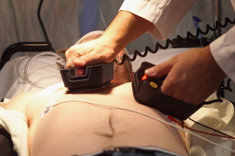
Electrical cardioversion uses electric shocks generated by the defibrillator and transmitted to the patient by means of electrodes applied to the chest.
Pharmacological cardioversion, on the other hand, involves the administration of specific anti-arrhythmic drugs.
Cardioversion is usually a planned treatment, which takes place in a hospital centre, but without hospitalisation.
In fact, at the end of the treatment, if everything has gone well, the patient can already be discharged and return home.
Electrical cardioversion is generally well tolerated even by elderly patients and is not dangerous
It is not contraindicated in patients with pacemakers or implantable defibrillators.
Contraindications are related to the full anaesthesia required for external electrical cardioversion, in order to spare the patient the pain and sensation of electric shock to the heart.
The risks of the procedure are minimal and complications rare; it can cause skin burns in the area where the electrodes are applied in the case of external electrical cardioversion and a temporary lowering of blood pressure.
An abnormal heart rhythm may occur following treatment.
If thrombi are present within the left atrium of the heart, they may detach and move to other districts following the shock, causing embolism.
For this reason, electrical cardioversion is preceded by a transesophageal echocardiogram and therapy with anticoagulant drugs.
THE RADIO OF RESCUERS AROUND THE WORLD? IT’S RADIOEMS: VISIT ITS BOOTH AT EMERGENCY EXPO
Performing Electrical Cardioversion
Scheduled electrical cardioversion is a procedure requiring admission to Day Hospital.
Before performing electrical cardioversion, the cardiologist informs the patient about the procedure and starts the preparation after signing the informed consent.
To avoid the twinges of pain caused by the electrical discharge, deep sedation with hypnoinducers will be performed, and in some cases, given the use of specific drugs, the anaesthetist will be called in.
Electric cardioversion involves the delivery of electric shocks with a defibrillator by means of two adhesive metal plates placed on the patient’s chest; these plates are positioned: right subclavear – left apical or antero-posterior.
Once sedation has been established, the cardiologist, adjusting himself according to the patient’s weight, will select the necessary discharge energy and synchronise the delivery of the shock with the electrocardiogram trend; the shock must be performed on the R peak because if it were to occur on the T wave it could cause the onset of malignant arrhythmias.
After ascertaining vital parameters, the doctor proceeds to deliver the shock; if the rhythm is not restored by the first shock, up to three shocks can be repeated by gradually increasing the joules.
The passage of electric current causes immediate contraction of the myocardial cells by resetting the abnormal circuits, allowing the restoration of sinus rhythm.
Restoration of normal cardiac rhythm occurs in 75-90% of cases in recent onset atrial fibrillation and 90-100% in cases of flutter arrhythmia.
Waking up the patient by monitoring his vital parameters
Convalescence after electrical cardioversion does not require any special precautions and you can return to your daily activities after 24 hours, unless otherwise indicated by your doctor.
It is necessary to carefully follow the prescribed maintenance therapy, be it anticoagulant drugs and, if necessary, anti-arrhythmic drugs.
In order to avoid relapses, it is useful to adopt a healthy lifestyle: reducing stress as much as possible, eliminating smoking and alcohol, and maintaining regular physical activity.
Read Also
Emergency Live Even More…Live: Download The New Free App Of Your Newspaper For IOS And Android
Difference Between Spontaneous, Electrical And Pharmacological Cardioversion
‘D’ For Deads, ‘C’ For Cardioversion! – Defibrillation And Fibrillation In Paediatric Patients
Inflammations Of The Heart: What Are The Causes Of Pericarditis?
Do You Have Episodes Of Sudden Tachycardia? You May Suffer From Wolff-Parkinson-White Syndrome (WPW)
Knowing Thrombosis To Intervene On The Blood Clot
Patient Procedures: What Is External Electrical Cardioversion?
Increasing The Workforce Of EMS, Training Laypeople In Using AED
Heart Attack: Characteristics, Causes And Treatment Of Myocardial Infarction
Altered Heart Rate: Palpitations
Heart: What Is A Heart Attack And How Do We Intervene?
Do You Have Heart Palpitations? Here Is What They Are And What They Indicate
Palpitations: What Causes Them And What To Do
Cardiac Arrest: What It Is, What The Symptoms Are And How To Intervene
Electrocardiogram (ECG): What It Is For, When It Is Needed
What Are The Risks Of WPW (Wolff-Parkinson-White) Syndrome
Heart Failure: Symptoms And Possible Treatments
What Is Heart Failure And How Can It Be Recognised?
Inflammations Of The Heart: Myocarditis, Infective Endocarditis And Pericarditis
Quickly Finding – And Treating – The Cause Of A Stroke May Prevent More: New Guidelines
Atrial Fibrillation: Symptoms To Watch Out For
Wolff-Parkinson-White Syndrome: What It Is And How To Treat It
Do You Have Episodes Of Sudden Tachycardia? You May Suffer From Wolff-Parkinson-White Syndrome (WPW)
What Is Takotsubo Cardiomyopathy (Broken Heart Syndrome)?
Heart Disease: What Is Cardiomyopathy?
Inflammations Of The Heart: Myocarditis, Infective Endocarditis And Pericarditis
Heart Murmurs: What It Is And When To Be Concerned
Broken Heart Syndrome Is On The Rise: We Know Takotsubo Cardiomyopathy
Heart Attack, Some Information For Citizens: What Is The Difference With Cardiac Arrest?
Heart Attack, Prediction And Prevention Thanks To Retinal Vessels And Artificial Intelligence
Full Dynamic Electrocardiogram According To Holter: What Is It?
In-Depth Analysis Of The Heart: Cardiac Magnetic Resonance Imaging (CARDIO – MRI)
Palpitations: What They Are, What Are The Symptoms And What Pathologies They Can Indicate
Cardiac Asthma: What It Is And What It Is A Symptom Of


