Causes, remedies and exercises for eyelid ptosis
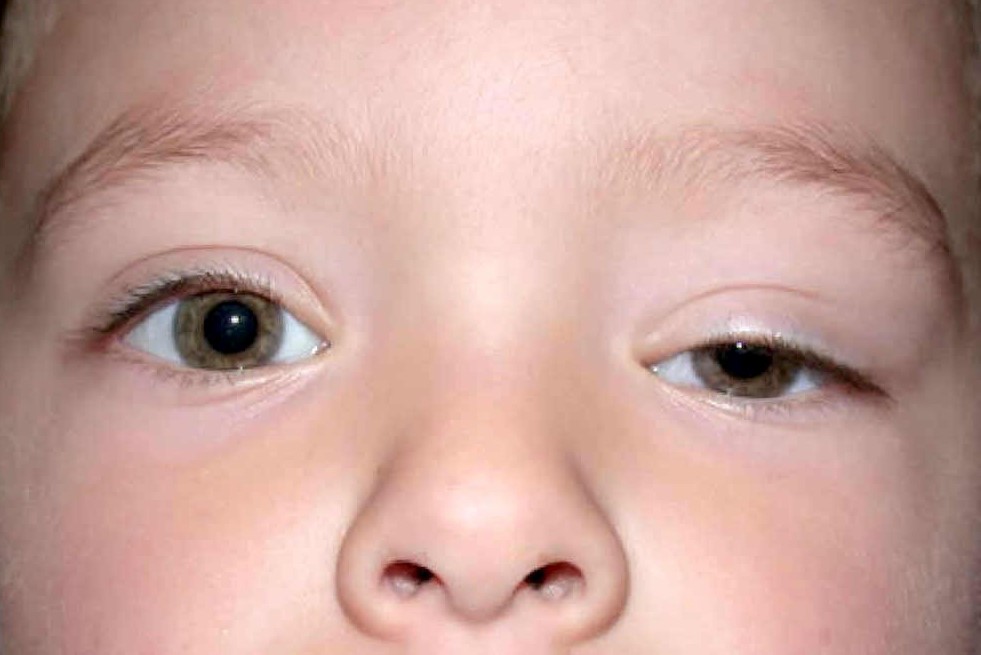
The term eyelid ptosis denotes a situation characterised by a drooping of the upper eyelid that, under normal conditions, laps at the upper edge of the cornea
Types of eyelid ptosis
From a normo-functional point of view, there are 3 forms of eyelid droop:
- slight: the drooping slightly exceeds the corneal edge;
- medium: the drooping is around the pupil;
- severe: the centre of the pupil is exceeded or, in some cases, covered completely.
Mild and medium forms are mainly a cosmetic problem, while severe forms prevent light from penetrating into the eye and this causes amblyopia in children and non-vision in adults.
The causes of eyelid ptosis
The causes of eyelid ptosis vary depending on the type, whether congenital or acquired.
Congenital eyelid ptosis is almost always bilateral and originates on the basis of a malformation affecting the eyelid suspension apparatus or the neurological component, leading to congenital alterations in the innervation of muscle structures.
The problem with congenital ptosis is that, if it is severe, it causes amblyopia in children, preventing them from developing proper visual function.
CHILD HEALTH: LEARN MORE ABOUT MEDICHILD BY VISITING THE BOOTH AT EMERGENCY EXPO
Acquired forms of eyelid ptosis, on the other hand, are often unilateral and can be determined by a cause
- neurogenic (of the nerves): they arise following ischaemia and are due to paralysis of the third cranial nerve that innervates the elevator muscle of the upper eyelid and also some muscles of the eye. The diagnosis is extremely easy in these cases: the eyelid is drooping and the underlying eye is completely squinting;
- myogenic (of the muscles): these are often bilateral and are favoured by myasthenia;
- mechanical: in this case, a voluminous neoformation of the upper eyelid such as, for example, an angioma, can lower the eyelid, just as a scar following trauma can lead to alterations in the eyelid muscles.
Lastly, there is senile ptosis favoured by the physiological involution of the tendon of the eyelid elevator muscle and due to enophthalmos, the abnormal sunken eyeball in the orbit, typical of the third age, which produces a retraction of the eyeball and, at the same time, a consequent lowering of the upper eyelid.
How it is treated
In severe forms of ptosis, surgery should be performed as soon as possible. In children, however, it is important to wait until the child’s body is fully developed before proceeding.
In any case, it is a complex operation during which the amount of muscle reinforcement to be inserted to restore proper adjustment must be determined very precisely.
If, in fact, the muscles are not strengthened well, the risk is an insufficient elevation of the eyelid, which in fact does not solve the problem, but if they are strengthened too much, there is an excessive elevation so that the eye no longer closes, exposing the patient to the risk of recurring ulcers and corneal perforations.
Medium or mild forms of ptosis, precisely because of the complexity of the surgical procedure, are not treated even though they represent a very significant aesthetic discomfort, in addition to causing an incorrect head position due to the desire to compensate for this lowering of the eyelid, which gives rise to an inevitable visual disturbance.
Eyelid ptosis exercises: are they effective?
In principle, exercises and eyelid gymnastics are of little use: in the presence of ptosis, in fact, either nothing is done or surgery is performed in order to restore the correct eyelid muscle balance.
In patients with paralysis and subsequent eyelid ptosis, so-called suspension surgery can be used, whereby the eyelid is hooked onto the frontalis muscle.
Eyelid ptosis and blepharocalasis: what is the difference
Many people confuse eyelid ptosis with blepharocalasis, i.e. the physiological relaxation of eyelid tissue, but they are 2 different clinical conditions.
With advancing age, many patients have an overabundance of skin on the eyelid: this skin forms a plica, a fold, that overlies the eyelid edge, simulating a ptosis.
The difference, however, is that in these cases it is not the eyelid edge that is lowered, but actually this excess skin that falls over the edge and can be effectively removed with a blepharoplasty.
Read Also:
Emergency Live Even More…Live: Download The New Free App Of Your Newspaper For IOS And Android
Pupillary Reflex To Light: Mechanism And Clinical Significance
4 Reasons To Seek Emergency Care For Vision Symptoms
Autoimmune Diseases: The Sand In The Eyes Of Sjögren’s Syndrome
Blepharoptosis: Getting To Know Eyelid Drooping
Corneal Abrasions And Foreign Bodies In The Eye: What To Do? Diagnosis And Treatment
Corneal Abrasions And Foreign Bodies In The Eye: What To Do? Diagnosis And Treatment
Wound Care Guideline (Part 2) – Dressing Abrasions And Lacerations
Contusions And Lacerations Of The Eye And Eyelids: Diagnosis And Treatment
Macular Degeneration: Faricimab And The New Therapy For Eye Health
First Aid For Dehydration: Knowing How To Respond To A Situation Not Necessarily Related To The Heat
Hydration: Also Essential For The Eyes
What Is Aberrometry? Discovering The Aberrations Of The Eye
Red Eyes: What Can Be The Causes Of Conjunctival Hyperemia?
What Is Corneal Abrasion And How To Treat It
Covid, A ‘Mask’ For The Eyes Thanks To Ozone Gel: An Ophthalmic Gel Under Study
Opening The Eyes Of The World, CUAMM’s “ForeSeeing Inclusion” Project To Combat Blindness In Uganda
What Is Ocular Pressure And How Is It Measured?
Blepharoptosis: Getting To Know Eyelid Drooping


