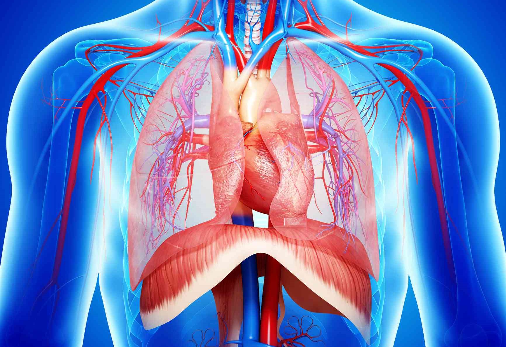
Chest trauma: traumatic rupture of the diaphragm and traumatic asphyxia (crushing)
There are several specific injuries that can occur in the setting of chest trauma, these commonly occur in combination with other more common injuries and can complicate the presentations of patients
The ones discussed here are diaphragmatic rupture, bronchial rupture, and traumatic asphyxia.
Types of chest trauma: Diaphragmatic Rupture
Rupture of the diaphragm can result in the abdominal contents crossing the base of the thorax/top of the abdomen and taking up space in the thoracic cavity.
This will often mimic the initial presentation of hemothorax or pneumothorax.
The most common cause of this injury is motor vehicle accidents, with penetrating trauma of the abdomen as a distant second.
The expected respiratory distress is often paired with abdominal tenderness and in some cases, the presence of bowel sounds in the thoracic cavity.
Often this is difficult to detect in a field environment and the injury will be mistaken for a pneumo/hemothorax.
The left side is more commonly affected due to the position of the liver on the right preventing intestines from traversing the ruptured diaphragm.
MANAGEMENT:
Management of diaphragmatic rupture is focused on providing 100% oxygen via a non-rebreather mask, placing large-bore IV’s, and treating any developing hypovolemia/hypoxemia.
Frequent re-assessment for the development of acute blood loss or increasing respiratory difficulty is vital to management as patients may rapidly decompensate due to difficulty ventilating or due to rapid blood loss.
Chest Trauma With Bronchial Rupture
Bronchial rupture commonly results from penetrating injury and rarely from severe blunt force trauma.
Any mechanism of injury significant enough to cause direct damage to the bronchial tree often also damages the surrounding lung and pleura.
This results in a complex injury that shares elements of hemothorax, pneumothorax, alveolar hemorrhage, and structural damage to the chest.
Patients with bronchial rupture will often be in respiratory distress that is minimally responsive to oxygen due to interruption of the airway and hypovolemia that results from significant blood loss into the tissue of the lung.
On assessment these patients will often have “crunchy” or harsh breath sounds over the area of injury as the damaged bronchus collapses and re-opens with each breath cycle.
Stridor, a high pitched whistling, is also possible if the larger mainstem bronchi and/or trachea is damaged.
MANAGEMENT: Management is focused on obtaining control of the damaged airway and preventing loss of blood/air into the tissue of the lung and the thoracic cavity that surrounds it.
Gain control of the airway by attempting to get an endotracheal tube distal to the damaged segment. Often this means intubating a mainstem bronchus.
The pressure from the inflated ET tube balloon at the end of the tube can help reduce bleeding by providing direct compression to the wound.
Limit compromise of breathing and ventilation by providing 100% oxygen via non-rebreather and gain control of circulation by placing large bore IV’s and providing IV fluids in reasonable amounts (generally 20 ml/kg total in 500ml boluses) to prevent profound hypotension below 90mmhg systolic.
Iatrogenic (from the intervention) harm is common in these patients. Intubation may contribute to tension pneumothorax by increasing the pressure of air going into the lung.
Excess blood filling the area distal to the bronchus can cut off entire areas of the lung, leading to hypoxia, and inadequate ventilation of the undamaged lung can also contribute to hypoxia.
Needless to say, these patients are critically ill and require continuous re-assessment while en route to a trauma facility.
Traumatic Asphyxia
Traumatic Asphyxia is a dramatic and hyper-acute syndrome that presents after a significant chest trauma temporarily disrupts cardiopulmonary mechanics.
Sudden and severe crushing forces on the chest results in the reversal of blood flow (reflux) from the right side of the heart back through the superior vena cava and into the large veins of the neck and head.
This can result in near-instant change in mental status, seizure, or new neurologic deficit. Seconds to minutes after bilateral subconjunctival hemorrhage, edema, swelling of the tongue, a bright red face, and upper extremity cyanosis commonly develop.
MANAGEMENT: Management is supportive unless other co-dominant injuries are present.
This condition is dramatic and alarming but rarely life-threatening.
As such a full assessment for thoracic, abdominal, spinal, and cranial injuries is warranted.
If indicated due to changes in vital signs or patient presentation initiate treatment with oxygen, IV fluids, and place the patient on monitors.
Read Also:
Emergency Live Even More…Live: Download The New Free App Of Your Newspaper For IOS And Android
Tracheal Intubation: When, How And Why To Create An Artificial Airway For The Patient
What Is Transient Tachypnoea Of The Newborn, Or Neonatal Wet Lung Syndrome?
Traumatic Pneumothorax: Symptoms, Diagnosis And Treatment
Diagnosis Of Tension Pneumothorax In The Field: Suction Or Blowing?
Pneumothorax And Pneumomediastinum: Rescuing The Patient With Pulmonary Barotrauma
ABC, ABCD And ABCDE Rule In Emergency Medicine: What The Rescuer Must Do
Multiple Rib Fracture, Flail Chest (Rib Volet) And Pneumothorax: An Overview
Internal Haemorrhage: Definition, Causes, Symptoms, Diagnosis, Severity, Treatment
Cervical Collar In Trauma Patients In Emergency Medicine: When To Use It, Why It Is Important
KED Extrication Device For Trauma Extraction: What It Is And How To Use It
How Is Triage Carried Out In The Emergency Department? The START And CESIRA Methods
Chest Trauma: Clinical Aspects, Therapy, Airway And Ventilatory Assistance


