Coccygodynia: definition, symptoms, diagnosis and treatment
Coccygodynia is a medical condition affecting the pelvis, particularly the coccyx, characterised by the presence of pain (frequently caused by inflammation) of the sacral area
It may occur as a result of trauma or may be due to other factors, such as postural defects or impaired mobility of the coccyx.
This disorder can affect both male and female patients, however, it has a higher incidence in women, while the average age of onset is usually around 40 years.
Although coccygodynia can be a source of discomfort and pain of varying intensity, it is generally a non-serious condition that poses no particular risk to the patient.
Nevertheless, it is important to exclude the presence of other associated pathologies.
Depending on the cause of the disorder and the nature of the pain, it is possible to opt for different therapeutic approaches, which may include pharmacological treatment to combat inflammation, physiotherapeutic manipulations to control the pain, or surgery in more severe cases.
What is Coccygodynia?
Coccygodynia is a syndrome affecting the pelvis and sacral area, resulting in severe chronic pain.
The word, in fact, is etymologically composed of coccyx, i.e. the bone at the end of the spinal column, and dinia, i.e. pain.
It is a widespread disorder among the population and can be quite disabling for the sufferer. Persistent pain in the coccyx prevents the patient from sitting or standing for long periods of time, limiting most daily activities.
Although coccygodynia can affect individuals of all ages and both sexes, it has a higher incidence in women and tends to occur mainly in adulthood.
The causes of the disorder are numerous: they may be traumatic in nature, may be related to other pathological disorders, or may be due to other factors such as stress, repetition of certain sports and work activities, overweight conditions, or even childbirth.
Anatomy of the coccyx
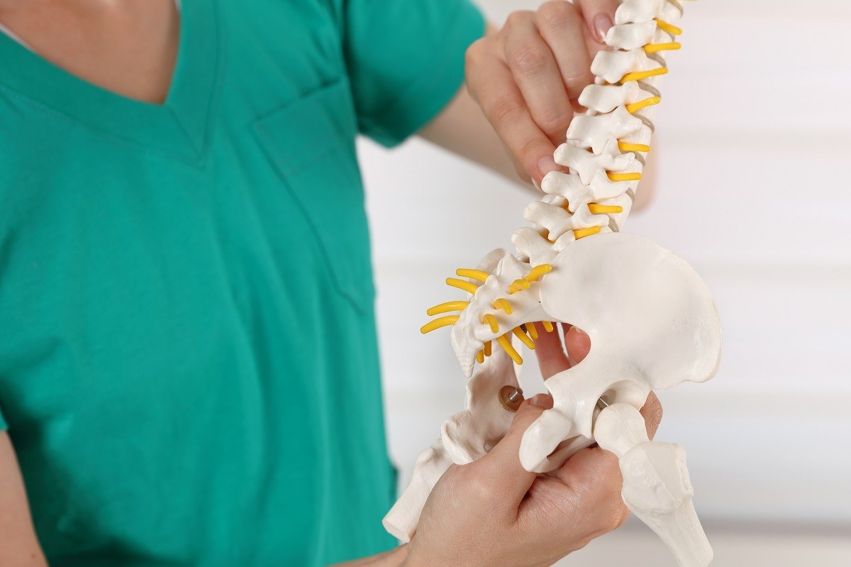
The coccyx is a small, triangular-shaped bone at the base of the sacrum, i.e. the pelvic weight-bearing bone, just above the cleft of the buttocks; it consists of 3 to 5 vertebral units that are called ‘false’ because, with the exception of the first segment, they do not have the typical characteristics of vertebrae and are fused together.
The coccyx can be divided into six segments: the base, the apex, the anterior area, the posterior area and the two lateral areas.
This bone has a slightly downward arched shape, with the apex of the terminal apex oriented towards the front of the body, reminiscent of a tail sketch that was probably present in earlier evolutionary stages of man.
Near the apex of the coccyx is the anal sphincter, while on the dorsal surface are the grafts for the gluteus maximus muscle, the anococcygeal ligament and the pubococcygeal muscle.
From an anato-functional point of view, the coccyx contributes to the protection of the spinal canal that ends in the lumbar spine.
In addition, it contributes to supporting the weight of the body and enables one to assume a sitting position.
Sometimes, due to postural vices, pathologies or other physiological factors, the coccyx can assume an incorrect position or inclination, causing pain and discomfort both at rest and when performing certain activities.
Coccygodynia, what can be the triggers?
As mentioned above, coccygodynia is frequently caused by chronic inflammation in the coccygeal area.
The triggering causes can be manifold: in most cases, accidents or traumatic events caused by impact of the coccyx with hard surfaces, or spinal trauma and falls are at the origin of the condition.
Other risk factors may be overloading of the lumbar region, childbirth, overweight conditions, or age-related wear and tear.
When no apparent cause can be identified, we speak of idiopathic forms.
Certain sporting activities, such as contact sports, skating, horse-riding or skiing, present a high risk of injury to the coccyx: although these are often simple contusions, violent trauma can also cause fractures and dislocations (i.e. displacement of the coccyx from its original anatomical position).
In these cases, the instability of the joint due to hypermobility of the coccygeal bones can trigger an inflammatory process that is the main cause of the pain and degeneration of the surrounding tissues typical of coccygodynia.
Coccygodynia may also occur as a consequence of repeated exertion involving high mobility of the sacro-coccygeal area, or as a result of poor posture while driving or working: continuous friction and persistent pressure on the lower vertebrae of the spinal column can induce microtrauma in the surrounding structures.
It is precisely for this reason that obese patients may experience posterior subluxation of the coccyx, as the heavy weight exerts greater intrapelvic pressure.
Another major cause of the onset of coccygodynia is tissue and cartilage degeneration due to ageing.
Pregnancy can also be considered a risk factor, as during the third trimester, hyperflexion of the coccyx occurs to facilitate delivery, which can lead to displacement of the joint.
More rarely, coccygodynia may be associated with the occurrence of tumours, osterosarcomas, pilonidal cysts, nerve root compression or infections.
What are the main symptoms?
Coccygodynia is a condition mainly characterised by localised pain or burning at the base of the back, where the coccyx is located.
The pain can vary from patient to patient and tends to worsen over time; in severe cases, it can become so intense that it impairs the performance of many daily activities, such as driving, sitting or stooping.
The discomfort tends to worsen especially when the patient changes from sitting to standing, or when assuming positions that put more pressure on the sacral area.
Patients with coccygodynia may also present with symptoms such as:
- Back pain and burning in the lumbar area;
- Pain in the legs, buttocks or hips;
- Constipation and pain before or during evacuation;
- Intestinal discomfort or stomach ache;
- Pain during the sexual act, although more rarely;
- Dysmenorrhoea in women, i.e. particularly painful menstrual cycle;
- In the case of injuries of traumatic origin, haematoma and bruising may occur;
Diagnosis
In general, a careful objective test by the doctor is sufficient to diagnose coccygodynia: he or she will assess the symptoms reported by the patient, collect a thorough personal and family history, and finally perform a thorough examination of the lower back region.
Through palpation, it will be possible to preliminarily exclude the presence of abnormal masses, abscesses and infections: as mentioned above, coccygodynia is not a particularly serious condition, but may be a symptom of other pathologies that need to be treated promptly.
To better frame the case under test, the patient should take an X-ray or MRI in both a sitting and standing position: comparing the images allows one to determine excessive or abnormal mobility of the sacro-coccygeal joint, which is itself quite rigid, and allows one to detect the presence of fractures and bone lesions.
Care and Treatment
The treatment of coccygodynia can vary greatly from case to case, depending on the underlying causes of the disorder and the extent of the reported symptoms.
In most cases, this disease responds well to targeted drug therapy based on:
- Analgesic drugs to reduce pain such as paracetamol and tramadol, or skin application of specific creams and ointments;
- Non-steroidal anti-inflammatory drugs (NSAIDs) that help reduce inflammation and have good analgesic properties. The most commonly used drugs include ibuprofen and naproxen;
- muscle relaxant drugs to relieve muscle contraction. An example would be thiocolchicoside;
For cases characterised by more intense symptoms, it is possible to opt for treatment with corticosteroid injections such as methylprednisolone or triamcinolone, together with local anaesthetics.
With this treatment approach, symptoms usually resolve within a few weeks.
In many cases, it may be useful to combine pharmacological treatments with a complementary therapy involving a period of rest and spinal manipulation techniques used in physiotherapy, osteopathy and chiropractic.
In addition, the use of special cushions and properly designed seats to relieve pressure on the coccyx can help provide pain relief when performing certain activities.
In more severe cases, when the conservative approach does not yield satisfactory results, it may be necessary to intervene with a surgical operation to resolve the disorder: coccygectomy consists of the total or partial removal of the coccyx vertebrae; in order to preserve all functionality, the muscles, tendons and ligaments that are attached to the removed bone segment are reconnected to the other parts of the pelvis.
Coccygectomy can require a rather long recovery time that can range from a few weeks to several months.
Read Also
Emergency Live Even More…Live: Download The New Free App Of Your Newspaper For IOS And Android
Coccygodynia: Symptoms, Diagnosis And Treatment
Ankylosing Spondylitis, What Are The Symptoms?
Chronic Pain And Psychotherapy: The ACT Model Is Most Effective
Lumbago: What It Is And How To Treat It
Back Pain: The Importance Of Postural Rehabilitation
Cervical Stenosis: Symptoms, Causes, Diagnosis And Treatment
Pain Therapy For Back Pain: How It Works
Prehospital Spine Immobilization In Penetrating Injuries: Yes Or No? What Do Studies Say?
Spinal Column Injuries, The Value Of The Rock Pin / Rock Pin Max Spine Board
Spinal Column Immobilisation Using A Spine Board: Objectives, Indications And Limitations Of Use


