Congenital diaphragmatic hernia (CHD): what it is, how to treat it
Congenital diaphragmatic hernia (CHD) is a malformation consisting of a lack of or incomplete formation of the diaphragm, i.e. a leakage of viscera from the abdomen (where they are normally located) into the thoracic cavity
Characteristics and consequences of congenital diaphragmatic hernia
The consequence is that the organs compress the lung on the herniated side, (in some cases also on the contralateral side), occupying space and preventing its normal development.
Severe and less severe forms are known, but the prognosis is usually severe, survival rates, with surgical resolution, vary worldwide between 50% and 70%.
This defect has a frequency of between 2,500 and 3,500 live births.
There is a slight predominance of males over females: 1 case per 3,000-5,000 live births.
It is not a hereditary disease although it has been described exceptionally in several individuals in the same family.
At present, the factors responsible for its formation are not fully known.
The diagnosis is usually made in the second trimester of pregnancy when the ultrasound scan shows one or more abdominal organs (intestine, spleen, stomach, liver).
The thorax and heart are generally displaced to the right, the left side being the most frequently herniated.
Diaphragmatic hernia, the picture in pregnancy
In the course of pregnancy, the ultrasound picture remains more or less stable, particular attention should be paid to the amniotic fluid, which may increase (polydramnios) causing premature birth.
It is important to bear in mind that: as long as the foetus is in the mother’s belly, it is not affected by poor lung development because it is the mother who provides it with nutrition and oxygenation.
Periodic check-ups (approx. every three to four weeks) are carried out to monitor the well-being of the foetus, assess the clinical picture and, if necessary, establish a treatment plan.
The medical team, following the diagnosis, has the task of providing the parents with all the information they need to make an informed decision, thus defining a shared course of treatment from the prenatal to the postnatal phase.
Antenatal diagnosis serves to ensure maximum care for the child, giving parents the opportunity to prepare for the experience and to establish a relationship with the medical team even before the child is born.
It would be advisable for parents to visit the ward where their baby will be received, cared for and treated, and to familiarise themselves with the people and the environment before the birth.
It is important for the baby to be born as late as possible; ideally after 38 weeks, and to be able to schedule the birth in a centre agreed upon with the team.
At birth, due to incomplete lung development, your baby will have significant breathing difficulties, which is why mechanical ventilatory assistance (intubation) must already be provided in the delivery room; the baby is then intubated and treated as soon as it is born.
The first 24/48 hours of a baby’s life are important to understand the degree of lung development achieved during foetal development.
The lungs are essential to life because they ensure gas exchange, i.e. they introduce oxygen and eliminate carbon dioxide.
Although you can give your baby plenty of oxygen, if he/she is unable to introduce it due to poor lung development, you will not be able to keep him/her alive.
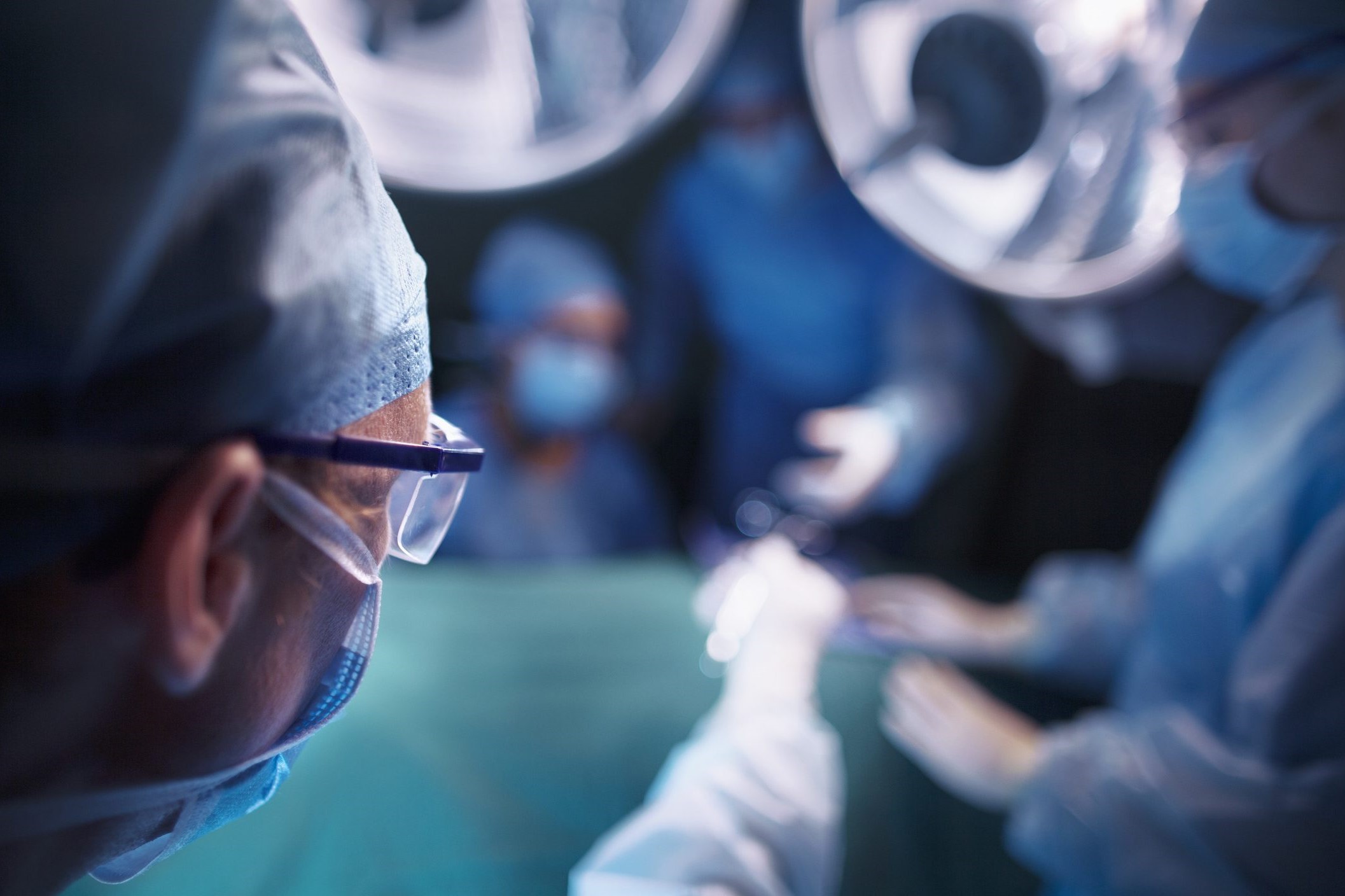
In some centres, surgery is carried out in the intensive care unit and not in the operating theatre, to avoid additional stress that can affect the child’s laboriously achieved respiratory stability The diaphragm defect (hernia) is corrected when the child is stable from a cardio-respiratory point of view.
Stable means that for a certain period of time the child needs the same amount of oxygen and the same type of ventilation, without any major fluctuations.
This can occur after 48 hours of life, if the child is able to ventilate stably, or a few days after birth.
The time needed to reach stabilisation is very variable and sometimes in very severe babies, it is never reached
The operation consists of making a subcostal incision (in the upper part of the abdomen), returning the herniated organs to the abdomen and reconstructing the integrity of the diaphragm.
If the diaphragm defect is large, it is necessary to use synthetic materials (diaphragmatic plate or patch).
In the days following surgery, the child, as long as he/she is intubated, will only be able to feed himself/herself through a nasogastric tube.
At the beginning the child may still be fatigued (shortness of breath) and may not be able to eat everything by mouth. In this case the help of the nasogastric tube will still be used for as long as he/she needs it.
It is useful for the mother to start expressing her milk immediately, so that she can breastfeed the baby and, in any case, give him/her her milk.
In the post-operative course, complications can occur, such as the persistence of a respiratory insufficiency that is difficult to treat and only slowly improves.
Infections, since one is operating on a newborn who has few immune defences and with numerous elements of further infectious risk Pleural effusion, which may require the placement of a chest drain.
Both before and after the operation, the presence of the parents is important for the child.
The first few weeks of life are by far the most difficult for both child and parents.
There is worry, hope, emotions all intense and conflicting. The parents’ role is fundamental, the doctors and nurses provide the necessary care, but tenderness, contact, attention, love, only the parents can guarantee these with their presence.
The duration of hospitalisation is very variable.
Each child has its own times and rhythms to learn and respect, without falling into the error of comparing them to others.
After a number of years, most children have good respiratory function as their lungs can recover to the point where they can live a normal life.
In other cases, children who have undergone diaphragmatic hernia surgery may present
- respiratory function problems, especially if intubation has been prolonged,
- gastro-oesophageal reflux problems,
- hearing problems of varying degrees and skeletal problems (scoliosis), which are more frequent when the plate was needed to reconstruct the diaphragm.
Read Also:
Emergency Live Even More…Live: Download The New Free App Of Your Newspaper For IOS And Android
What It Is And How To Recognise Abdominal Diastasis
Chronic Pain And Psychotherapy: The ACT Model Is Most Effective
Hiatal Hernia: What It Is And How To Diagnose It
Percutaneous Discectomy For Herniated Discs
What Is That Swelling? Everything You Need To Know About Inguinal Hernia
Symptoms And Causes Of Umbilical Hernia Pain


