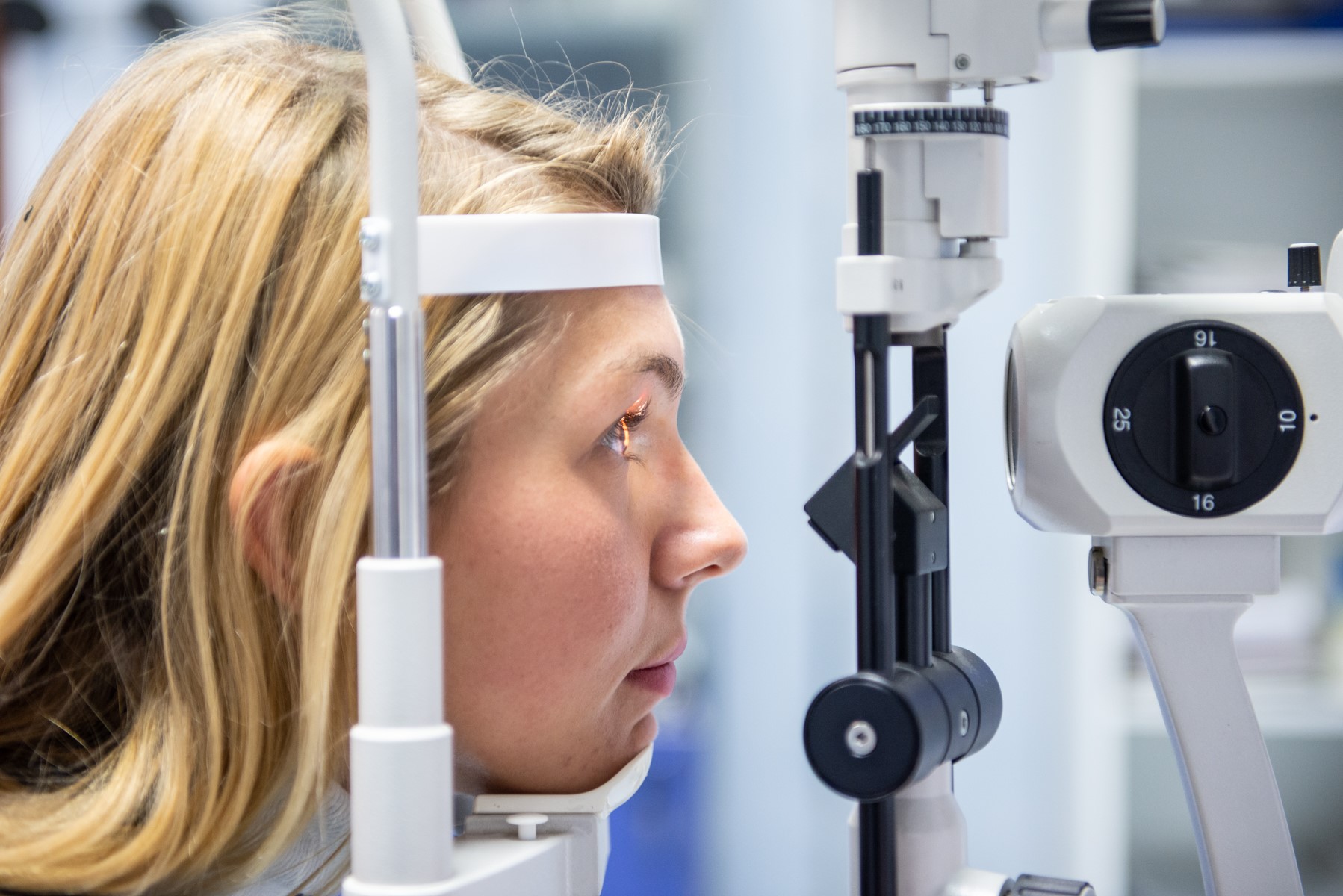
Do you suffer from diabetic retinopathy? Here's what's happening to you and what treatments are available
Let’s talk about diabetic retinopathy: diabetes that is not treated as it should be can, in the long run, trigger consequences in various areas of the body
This is the case with diabetic retinopathy, where hyperglycaemia damages the eye capillaries, which become weak and permeable.
Diabetic retinopathy is asymptomatic at its onset, but can degenerate by blurring vision, at first mildly, then evolving into blindness.
It affects both eyes and is more likely to develop in long-term diabetic patients.
Several patients complain of the first symptoms some ten years after the first diagnosis of diabetes.
To date, Italian estimates indicate that there are approximately 3 million diabetic patients, of whom as many as 2 million have developed retinal complications.
Diabetic retinopathy is one of the main causes of blindness during adulthood
For precisely these reasons, all diabetic patients are recommended to undergo an annual eye examination to prevent the progression of the disease from permanently affecting the visual organs.
Effects of hyperglycaemia on the retina
The eye is a very delicate and complex organ that, in order to function properly, uses various membranes and anatomical corpuscles, each with its own precise function.
The retina is its most functional and delicate area, since it is the only one capable of collecting light stimuli from the outside world and converting them into electrical impulses to be sent to the brain (through the optic channels) for processing into three-dimensional images.
In order to function properly, the retina also needs blood and oxygen, transported by the tiny capillaries located near its surface. That makes it easy to understand why hyperglycaemia, by damaging blood vessels throughout the body, can also weaken retinal vessels, leading to vision problems.
It is typical for diabetics to complain of blurred vision, directly caused by damaged capillaries.
High levels of glucose in the blood make the small blood vessels weaker and more permeable, causing fluid and lipids to leak out and deposit on the ocular fundus.
These deposits eventually lead to oedema and, later, to retinal ischaemia that permanently compromises the visus.
The first stage of diabetic retinopathy, the mildest, is called Non-Proliferative Diabetic Retinopathy (NPDR).
If this becomes chronic, Diabetic Retinopathy becomes Proliferative (PDR): to make up for the capillaries that are out of order, the body generates new ones, in a slow process of neovascularisation.
Types of diabetic retinopathy
The medical community has drawn up two different classifications of diabetic retinopathy, which correspond to the intensity with which the symptoms present.
We speak of Non-Proliferative Diabetic Retinopathy (NPDR) when the disease is in its early stages and symptoms are mild.
The ocular capillaries begin to appear as weakened, due to high blood glucose levels in the blood, which alter the permeability of their walls.
This paves the way for the formation of blood disorders such as small aneurysms, oedemas and thromboses that generate haemorrhages in the eye, impairing vision.
The first deposits of lipids from the blood, known as exudates, can also be created.
When NPDR evolves to a chronic stage, we are faced with the so-called Proliferative Diabetic Retinopathy (PDR), a more advanced condition of the disease, in which blood capillaries are almost or completely occluded, due to high lipid deposition.
The subject develops very worrying retinal ischemia, which further worsens the visual picture.
Since the supply of oxygenated blood to the retina and eyes in general is still necessary, the organism predisposes itself to neovascularisation, i.e. the formation of new blood vessels in the retina.
However, the new blood vessels are abnormal and fragile and can quickly lead to retinal detachment with haemovascularisation or to a high fluid release resulting in glaucoma.
Finally, there is a third small case history.
When the visual changes are so small as to be almost imperceptible and quietly resolvable, we speak of a simple or background retinopathy.
Symptoms
It is not always possible to identify and treat diabetic retinopathy at an early stage because, in many cases, the condition is asymptomatic.
The patient may not realise the real situation they are in until symptoms are already advanced and vision begins to be blurred.
The most frequent symptoms in cases of retinal diabetes are as follows (the list is not exhaustive and refers to both NPDR and PDR cases):
- Blurred vision and loss of visual acuity. Occlusions and bleeding occurring in the ocular capillaries literally obscure the retina.
- Visual field with obscured areas. This is also a consequence of occlusion of the retinal capillaries.
- Myodesopias. It is common that, in addition to blurred vision, the patient complains of the vision of black spots and threads floating in front of the eyes.
- Hypovision. In general, the subject complains of a visual deficit (i.e. sees less than before).
- Reduced ability to see in the dark.
- Difficulty perceiving and distinguishing colours.
- Blindness. This is the most serious situation, associated with already advanced retinal diabetes. The loss of sight is a major psychological problem for those affected, not only because one of the five senses is lost but also because, when it arrives, the loss is sudden and immediately severe.
The symptoms of diabetic retinopathy usually appear about ten years after the diagnosis of diabetes and increase with the natural progression of the disease.
Their intensity is more serious in individuals who have not treated their diabetes correctly for a long time.
Causes and risk factors
The main cause of the impairment of retinal capillaries is high blood glucose levels, which make their walls weaker and more permeable, allowing liquids and lipids to pass freely and deposit on the ocular fundus.
Generally, this happens when diabetes has been present for many years and the right steps have not been taken to treat it.
After 15 to 20 years of such a condition, 80 per cent of individuals develop diabetic complications in both eyes.
Actively intervening in blood glucose contrast means slowing the rate of onset and progression of any diabetic complication, including retinal complications.
Controlling blood pressure is crucial. If an individual is hypertensive, his or her blood vessels are already stressed and compromised. Constant blood pressure control also has a beneficial effect on the progression of diabetic retinopathy.
High blood lipid levels, such as cholesterol and triglycerides, lead to an accumulation of exudates in the retina. Deposits form that obstruct the retina’s small blood vessels, impairing vision.
Pregnancy can also be an important cause of diabetic retinopathy, due to the major hormonal changes taking place, which can affect blood sugar levels. However, the progression of the disease often comes to a halt after childbirth.
Diagnosis of dibetic retinopathy
The road to the diagnosis of diabetic retinopathy passes through a specialist examination by an ophthalmologist.
During the anamnesis phase, it will be his or her task to collect the patient’s symptoms and clinical history, in order to prepare the most suitable subsequent tests and treatments.
The objective test, aimed at investigating the true stage of severity of the disease, is carried out using a special instrument called a retinograph, which, as its name suggests, carefully observes the ocular fundus, showing the state of health of the retina.
It is also useful to see how long the disease has been affecting the health of the retina.
Fluorangiography is a further technique used when the aim is to detect retinal microaneurysms and ischaemia. It assesses the extent of the disease by injecting a dye called fluorescein into the blood vessels, which highlights changes in the capillaries.
Finally, the ocular CT scan, known as Optical Coherence Tomography, observes in detail the macula and optic nerve, i.e. the two parts of the retina that are indispensable for collecting stimuli and rendering three-dimensional images. The typical light beam of the CT scan highlights any retinal lesions and fluid and lipid effusions in this area.
In the case of diabetic retinopathy, early diagnosis is essential in order to intervene immediately.
This is why diabetic patients should undergo an annual ophthalmic test.
Pregnant diabetic patients should be kept under observation, as the possibility of developing retinopathy increases.
Effective treatment and prevention
There are various types of treatment that are more or less effective depending on the type of diabetic retinopathy in progress (NPDR or PDR).
They can sometimes be used in combination with each other.
Therapies for NPDR (Non-Proliferative Diabetic Retinopathy)
Non-Proliferative Diabetic Retinopathy can be relieved by laser photocoagulation of the retina, a particularly innovative technique that uses the power of the laser to reduce swelling in the retina and macula.
Although it does not eliminate the condition, it certainly decreases the rate of disease progression, and restores relief and visual acuity. It also prevents serious complications such as haemvitreous and glaucoma.
Eyes with diabetic retinopathy can be treated with intravitreal injections.
Again, the injected drugs, which are completely safe, act to eliminate macular oedema and decrease the threshold of neovascularisation, restoring the individual’s normal vision.
NPDR can also be treated with the technique of photoablation, i.e. laser removal of the small damaged part of the cornea and retina.
Therapies for PDR
When diabetic retinopathy has reached its advanced and proliferating stage, the two most effective methods for maintaining as optimal a visual condition as possible are intraocular corticosteroid injections and vitrectomy.
While the former, thanks to the action of cortisone, significantly reduces pain and retinal oedema, the latter is a special surgery performed when there is a retinal detachment and consequent haemovitreous.
It serves to restore normal vitreous function without deposits of blood and other substances hindering it.
Generally, following this operation, vision improves greatly compared to the initial situation.
Implementing prevention strategies for diabetic retinopathy is not easy because the disease is often asymptomatic in its early stages.
It goes without saying that it is important to undergo constant eye check-ups, especially in long-term diabetics.
Continuous measurements of blood glucose and blood pressure values should not be missing in preventive treatment.
Read Also
Emergency Live Even More…Live: Download The New Free App Of Your Newspaper For IOS And Android
Diabetic Retinopathy: The Importance Of Screening
Diabetic Retinopathy: Prevention And Controls To Avoid Complications
Diagnosis Of Diabetes: Why It Often Arrives Late
Diabetic Microangiopathy: What It Is And How To Treat It
Diabetes: Doing Sport Helps Blood Glucose Control
Type 2 Diabetes: New Drugs For A Personalised Treatment Approach
Diabetes And Christmas: 9 Tips For Living And Surviving The Festive Season
Diabetes Mellitus, An Overview
Diabetes, Everything You Need To Know
Type 1 Diabetes Mellitus: Symptoms, Diet And Treatment
Type 2 Diabetes Mellitus: Symptoms And Diet
Semaglutide For Obesity? Let’s See What The Anti-Diabetic Drug Is And How It Works
Italy: Semaglutide, Used For Type 2 Diabetes, Is In Short Supply
Gestational Diabetes, What It Is And How To Deal With It
Diabesity: What It Is, What Risks And How To Prevent It
Wounds And Diabetes: Manage And Accelerate Healing
The Diabetic Diet: 3 False Myths To Dispel
Top 5 Warning Signs Of Diabetes
Signs Of Diabetes: What To Look Out For
What Is Presbyopia And When Does It Occur?
False Myths About Presbyopia: Let’s Clear The Air
Droopy Eyelids: How To Cure Eyelid Ptosis?
What Is Ocular Pterygium And When Surgery Is Necessary
Tear Film Dysfunction Syndrome, The Other Name For Dry Eye Syndrome
Vitreous Detachment: What It Is, What Consequences It Has
Macular Degeneration: What It Is, Symptoms, Causes, Treatment
Conjunctivitis: What It Is, Symptoms And Treatment
How To Cure Allergic Conjunctivitis And Reduce Clinical Signs: The Tacrolimus Study


