Echocardiogram: what it is and when it is required
The echocardiogram is a technique that is based on the use of ultrasound and makes it possible to see from the inside both the walls of the heart and its internal structures (such as the valves) and to accurately assess the overall contractility of the heart
It is a diagnostic technique that makes it possible to check specific areas of the heart muscle, the thickness and thickening of the walls, and their motility during cardiac activity.
It is a non-invasive, painless and uncomfortable test that does not require any specific preparation.
When it is necessary to perform an echocardiogram
The echocardiogram provides valuable information both in the acute phase of an infarction and in the post-infarction period.
Ischaemia and infarction induce evident alterations, transitory or stable respectively, in the motility of the regions of the heart muscle affected by the process: the echocardiogram makes it possible to confirm or not the diagnosis of infarction or ischaemia and to assess the exact site and extent of the damage, as well as to monitor the alterations highlighted and their possible changes over time.
The information that can be obtained with the echocardiogram has been increased by the possibility of performing the test in association with various types of stimulation aimed at better highlighting the infarcted or ischaemic areas of the muscle, and which essentially include physical exertion (stress echocardiogram) or the infusion during the test of particular drugs.
Nowadays, it is possible to check the heart as early as the first weeks of a foetus’ life and detect any malformations that need to be corrected with interventions after birth.
A new technique, trans-abdominal ultrasound, makes it possible, as early as the first trimester of pregnancy, to clearly show the anatomical structures of the heart, whereas previously one had to wait until the 5th or 6th month.
The non-invasive diagnosis of congenital heart disease plays an important role in the diagnostic process of the child with congenital heart disease.
The echocardiogram is an absolutely harmless and repeatable test
This diagnostic possibility is important above all to reassure couples who, due to hereditary factors, are at a higher risk of having a child with cardiac malformations or those who have already had a child with this problem.
Early diagnosis allows early control and paves the way for the first prenatal interventions that have recently been carried out.
What is the transesophageal echocardiogram
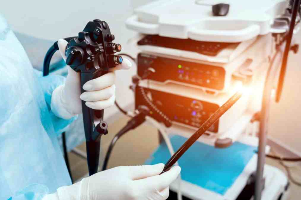
The transesophageal echocardiogram uses a probe that emits ultrasound.
The probe is passed through the mouth through the pharynx to the oesophagus and by exploiting the anatomical proximity of the posterior wall of the heart to the oesophagus provides better image quality than the transthoracic echocardiogram.
The test requires anaesthesia to be performed.
Read Also
Emergency Live Even More…Live: Download The New Free App Of Your Newspaper For IOS And Android
Fusion Prostate Biopsy: How The Examination Is Performed
What Is Needle Aspiration (Or Needle Biopsy Or Biopsy)?
What Is Echocolordoppler Of The Supra-Aortic Trunks (Carotids)?
What Is The Loop Recorder? Discovering Home Telemetry
Cardiac Holter, The Characteristics Of The 24-Hour Electrocardiogram
Peripheral Arteriopathy: Symptoms And Diagnosis
Endocavitary Electrophysiological Study: What Does This Examination Consist Of?
Cardiac Catheterisation, What Is This Examination?
Echo Doppler: What It Is And What It Is For
Transesophageal Echocardiogram: What Does It Consist Of?
Venous Thrombosis: From Symptoms To New Drugs
Echotomography Of Carotid Axes
Echo- And CT-Guided Biopsy: What It Is And When It Is Needed
Echodoppler: What It Is And When To Perform It


