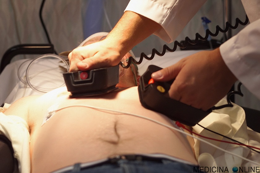
Electrical cardioversion: what it is, when it saves a life
Electrical cardioversion, CVE, is a therapeutic procedure used to restore normal heart rhythm in patients with atrial fibrillation, flutter, or tachycardia and in whom pharmacological cardioversion has failed
The most common cause of this type of abnormality is heart disease
Sometimes the patient perceives the alteration, but often he only notices the consequences it entails, such as palpitations, weakness, dizziness, fainting, asthenia.
The high heart rate caused by these arrhythmias damages the myocardial muscle as, if persistent, they lead to a reduction in contractile function and a reduction in the ejection fraction; ejection fraction which allows to evaluate the efficacy of the pump function of the heart and represents a good indicator of myocardial contractility.
In the case of atrial fibrillation, the lack of contractility in the atria causes abnormal blood circulation in the heart cavities, and in arrhythmias that last for more than 48 hours, thrombi can form in some parts of the atrium; thrombi which, following the resumption of atrial contractility, could fragment and disperse in the arterial circulation causing stroke and/or embolism.
An accurate anamnesis on the timing of the onset of symptoms plays a decisive role on the therapy to be adopted; if more than 48 hours pass from the onset of symptoms, it is mandatory to undertake a period of anticoagulant therapy at the end of which it is possible to carry out electrical cardioversion safely, minimizing the cardio-embolic risks.
There are two types of cardioversion, electrical and pharmacological
- Electrical cardioversion makes use of electrical discharges, shocks, generated by the defibrillator and transmitted to the patient via electrodes applied to the chest.
- Pharmacological cardioversion, on the other hand, involves the administration of specific antiarrhythmic drugs.
Cardioversion is usually a scheduled treatment that takes place in a hospital setting, but without hospitalization.
In fact, at the end of the therapy, if everything has gone well, the patient can already be discharged and go home.
Electrical cardioversion is generally well tolerated even by elderly patients and is not dangerous
It is not contraindicated in patients with pacemakers or implantable defibrillators.
The contraindications are related to the general anesthesia required for external electrical cardioversion, to avoid the pain and sensation of an electric shock to the heart for the patient.
The risks of the procedure are minimal and the complications rare; may cause skin burns in the area where the electrodes were applied in the case of external electrical cardioversion and a temporary drop in blood pressure. An abnormal heart rhythm may develop following treatment.
If there are thrombi inside the left atrium of the heart, following the shock they could detach and move to other districts, causing embolisms.
For this reason, electrical cardioversion is preceded by the execution of a transesophageal echocardiogram and by therapy with anticoagulant drugs.
Carrying out electrical cardioversion
Programmed electrical cardioversion is a procedure that requires day hospital admission.
Before carrying out the electrical cardioversion, the cardiologist informs the patient about the procedure and begins the preparation after signing the informed consent.
To avoid the pangs of pain due to the electric discharge, deep sedation with hypnoinducing will be performed, in some cases, given the use of specific drugs, the anesthetist will be used.
Electric cardioversion involves the delivery of electric shocks with a defibrillator using two adhesive metal plates placed on the patient’s chest; plates to be positioned: right subclavicular – left apical or anterior – posterior.
Once sedation has been ascertained, the cardiologist, adjusting on the basis of the patient’s weight, will select the necessary discharge energy and synchronize the delivery of the shock with the progress of the electrocardiogram; shock that must be performed on the R peak because if it occurred on the T wave it could cause the onset of malignant arrhythmias.
After ascertaining the vital parameters, the doctor proceeds with the delivery of the shock; if the rhythm does not restore with the first shock, up to 3 shocks can be repeated, gradually increasing the Joules.
The passage of electric current determines the immediate contraction of the myocardial cells by resetting the abnormal circuits, allowing the restoration of the sinus rhythm.
Restoration of normal heart rhythm occurs in 75-90% of cases in recent onset atrial fibrillation and in 90-100% in case of flutter arrhythmia. The patient will be awakened by monitoring his vital parameters.
Convalescence after electrical cardioversion does not require particular precautions and you can return to daily activities after 24 hours, unless otherwise indicated by your doctor.
It is necessary to carefully follow the prescribed maintenance therapy, both anticoagulant drugs and, if necessary, anti-arrhythmic drugs.
In order to avoid relapses it is useful to adopt a healthy lifestyle: reducing stress as much as possible, eliminating smoking and alcohol, maintaining regular physical activity.
Read Also
Emergency Live Even More…Live: Download The New Free App Of Your Newspaper For IOS And Android
Cardiac Rhythm Restoration Procedures: Electrical Cardioversion
Difference Between Spontaneous, Electrical And Pharmacological Cardioversion
‘D’ For Deads, ‘C’ For Cardioversion! – Defibrillation And Fibrillation In Paediatric Patients
Inflammations Of The Heart: What Are The Causes Of Pericarditis?
Do You Have Episodes Of Sudden Tachycardia? You May Suffer From Wolff-Parkinson-White Syndrome (WPW)
Knowing Thrombosis To Intervene On The Blood Clot
Patient Procedures: What Is External Electrical Cardioversion?
Increasing The Workforce Of EMS, Training Laypeople In Using AED
Heart Attack: Characteristics, Causes And Treatment Of Myocardial Infarction
Altered Heart Rate: Palpitations
Heart: What Is A Heart Attack And How Do We Intervene?
Do You Have Heart Palpitations? Here Is What They Are And What They Indicate
Semeiotics Of The Heart And Cardiac Tone: The 4 Cardiac Tones And The Added Tones
Heart Murmur: What Is It And What Are The Symptoms?
Branch Block: The Causes And Consequences To Take Into Account
Cardiopulmonary Resuscitation Manoeuvres: Management Of The LUCAS Chest Compressor
Supraventricular Tachycardia: Definition, Diagnosis, Treatment, And Prognosis
Identifying Tachycardias: What It Is, What It Causes And How To Intervene On A Tachycardia
Myocardial Infarction: Causes, Symptoms, Diagnosis And Treatment
Aortic Insufficiency: Causes, Symptoms, Diagnosis And Treatment Of Aortic Regurgitation
Congenital Heart Disease: What Is Aortic Bicuspidia?
Atrial Fibrillation: Definition, Causes, Symptoms, Diagnosis And Treatment
Ventricular Fibrillation Is One Of The Most Serious Cardiac Arrhythmias: Let’s Find Out About It
Atrial Flutter: Definition, Causes, Symptoms, Diagnosis And Treatment
What Is Echocolordoppler Of The Supra-Aortic Trunks (Carotids)?
What Is The Loop Recorder? Discovering Home Telemetry
Cardiac Holter, The Characteristics Of The 24-Hour Electrocardiogram
Peripheral Arteriopathy: Symptoms And Diagnosis
Endocavitary Electrophysiological Study: What Does This Examination Consist Of?
Cardiac Catheterisation, What Is This Examination?
Echo Doppler: What It Is And What It Is For
Transesophageal Echocardiogram: What Does It Consist Of?
Paediatric Echocardiogram: Definition And Use
Heart Diseases And Alarm Bells: Angina Pectoris
Fakes That Are Close To Our Hearts: Heart Disease And False Myths
Sleep Apnoea And Cardiovascular Disease: Correlation Between Sleep And Heart
Myocardiopathy: What Is It And How To Treat It?
Venous Thrombosis: From Symptoms To New Drugs
Cyanogenic Congenital Heart Disease: Transposition Of The Great Arteries
Heart Rate: What Is Bradycardia?
Consequences Of Chest Trauma: Focus On Cardiac Contusion
Performing The Cardiovascular Objective Examination: The Guide


