Electrocardiography: what it is and how to do it
Electrocardiography, how important is it? Routine checks for our health are really very important
These certainly include the ECG, electrocardiography, or electrocardiogram, a diagnostic test that is absolutely painless and can tell us a lot about our state of health.
It is usually performed during scheduled check-ups or when some disturbance of the cardiovascular system is suspected.
Today, this test can identify the presence of disturbances in the heart rhythm and even prevent serious complications.
Electrocardiography, how it works
We are now used to calling it an electrocardiogram, a simple test that can prevent major problems in the cardiovascular system.
Electrocardiography is in fact neurophysiological monitoring that records the electrical activity inherent in cardiac contractions.
This electrical activity is recorded using an electrocardiograph.
The electrocardiograph is a diagnostic instrument, a kind of recorder that is connected to electrodes.
These are placed in the wrists and ankles and at six points on the chest in order to make a recording of cardiac activity and then transmit it to the device.
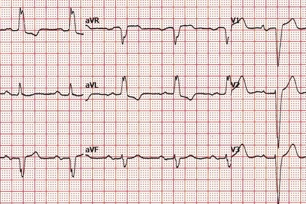
The electrical impulses detected are transcribed onto a sheet of graph paper at a rate of 25 mm per second, this tracing is called an electrocardiogram.
The electrocardiogram
Normally, the electrocardiogram consists, for each cardiac cycle, of a certain number of deflections in chronological succession followed by a period of cardiac rest, the diastoles.
This test is very simple and has no side effects; it can also be performed under stress, so we speak of dynamic electrocardiography, should the doctor deem it appropriate to perform this type of investigation as well.
Then there is a type of ECG which is instead performed in a more invasive manner.
By introducing a catheter into a vessel, the heart’s cardiac activity is recorded by inserting electrodes into it.
In this case, however, we are talking about endocavitary electrocardiography and it is a much less widely used variant of the ECG.
By means of the electrocardiogram, the condition of the heart muscle can be assessed and metabolic disorders can also be detected.
When is electrocardiography performed
Generally, electrocardiography is prescribed by one’s general practitioner along with other tests in order to ascertain possible problems, so it can be a routine test.
Regular check-ups are usually recommended after the age of 40 and may be more frequent with advancing age, but this is in the absence of any particular pathology.
If there is a family history of heart problems or if you are over 60, it is a good idea to ask your doctor how often to have check-ups in order to prevent cardiovascular disorders.
When electrocardiography is urgently needed
Unfortunately, there are conditions in which an electrocardiogram can be carried out with a certain urgency, such as when chest pain or various complaints are detected that may lead one to suspect a cardiological problem.
A chest pain is not a symptom of a heart attack, it could be a number of things, which is why it is a good idea, especially in certain situations, not to waste time and to expose the problem to the doctor.
What can be detected with the ECG
There are various pathologies affecting the cardiovascular system, many of which can be detected by ECG, let’s look at some of them:
- myocardial infarction: caused by a sudden interruption of blood flow to the heart;
- arrhythmias: if the heart beats too slowly, bradycardia, or if the heart beats too fast, tachycardia;
- ischaemic heart disease: interruption or blockage of the blood supply to the heart caused by narrowing of the coronary artery.
Types of ECG
As we have seen before there are different methods of performing this test, here are the main ones:
- Resting ECG: performed in a lying and supine position;
- Exercise ECG: performed during exercise of increasing intensity, on an exercise bike or treadmill;
- Dynamic ECG: performed with cardiac holter, then monitoring daily activity for a couple of days.
Read Also
Emergency Live Even More…Live: Download The New Free App Of Your Newspaper For IOS And Android
Electrocardiogram (ECG): What It Is For, When It Is Needed
What Are The Risks Of WPW (Wolff-Parkinson-White) Syndrome
Heart Failure: Symptoms And Possible Treatments
What Is Heart Failure And How Can It Be Recognised?
Inflammations Of The Heart: Myocarditis, Infective Endocarditis And Pericarditis
Quickly Finding – And Treating – The Cause Of A Stroke May Prevent More: New Guidelines
Atrial Fibrillation: Symptoms To Watch Out For
Wolff-Parkinson-White Syndrome: What It Is And How To Treat It
Do You Have Episodes Of Sudden Tachycardia? You May Suffer From Wolff-Parkinson-White Syndrome (WPW)
What Is Takotsubo Cardiomyopathy (Broken Heart Syndrome)?
Heart Disease: What Is Cardiomyopathy?
Inflammations Of The Heart: Myocarditis, Infective Endocarditis And Pericarditis
Heart Murmurs: What It Is And When To Be Concerned
Broken Heart Syndrome Is On The Rise: We Know Takotsubo Cardiomyopathy
Heart Attack, Some Information For Citizens: What Is The Difference With Cardiac Arrest?
Heart Attack, Prediction And Prevention Thanks To Retinal Vessels And Artificial Intelligence
Full Dynamic Electrocardiogram According To Holter: What Is It?
In-Depth Analysis Of The Heart: Cardiac Magnetic Resonance Imaging (CARDIO – MRI)
Palpitations: What They Are, What Are The Symptoms And What Pathologies They Can Indicate
Altered Heart Rate: Palpitations
Heart: What Is A Heart Attack And How Do We Intervene?
Do You Have Heart Palpitations? Here Is What They Are And What They Indicate
Palpitations: What Causes Them And What To Do
Cardiac Arrest: What It Is, What The Symptoms Are And How To Intervene
Cardiac Asthma: What It Is And What It Is A Symptom Of


