Emergency paediatrics / Neonatal respiratory distress syndrome (NRDS): causes, risk factors, pathophysiology
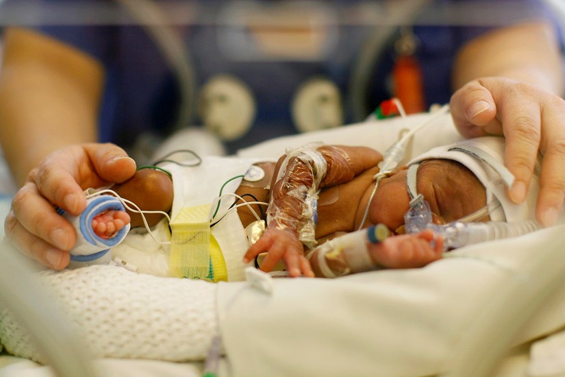
Neonatal respiratory distress syndrome (NRDS) is a respiratory syndrome characterised by the presence of progressive pulmonary atelectasis and respiratory failure that is mainly diagnosed in the premature neonate, who has not yet reached complete lung maturation and adequate surfactant production
Synonyms of infant respiratory distress syndrome are:
- ARDS of the infant (ARDS stands for acute respiratory distress syndrome);
- ARDS of the newborn;
- Neonatal ARDS;
- Paediatric ARDS;
- neonatal RDS (RDS stands for ‘respiratory distress syndrome’);
- newborn respiratory distress syndrome;
- acute respiratory distress syndrome of the child;
- acute respiratory distress syndrome of the newborn.
Respiratory distress syndrome was formerly known as ‘hyaline membrane disease’ hence the acronym ‘MMI’ (now fallen into disuse)
Infant respiratory distress syndrome in English is called:
- infantile respiratory distress syndrome (IRDS);
- respiratory distress syndrome of newborn;
- neonatal respiratory distress syndrome (NRDS);
- surfactant deficiency disorder (SDD).
The syndrome was previously known as ‘hyaline membrane disease’ hence the acronym ‘HMD’.
Epidemiology of newborn respiratory distress syndrome
The prevalence of the syndrome is 1-5/10,000.
The syndrome affects approximately 1% of newborns.
The incidence decreases with advancing gestational age, from about 50% in children born at 26-28 weeks to about 25% at 30-31 weeks.
The syndrome is more frequent in males, Caucasians, infants of diabetic mothers and second-born preterm twins.
Although there are many forms of respiratory failure affecting the newborn, NRDS is the predominant cause in the premature.
Advances in the prevention of premature birth and treatment of neonatal NRDS have led to a significant reduction in the number of deaths from this condition, although, NRDS continues to be a significant cause of morbidity and mortality.
It is estimated that approximately 50 per cent of newborn deaths have NRSD.
Because of the high mortality, all neonatal intensive care physicians should be able to diagnose and treat this common cause of respiratory failure.
Age of onset
The age of onset is neonatal: symptoms and signs of respiratory distress syndrome appear in the newborn soon after birth or a few minutes/hours after birth.
CHILD HEALTH: LEARN MORE ABOUT MEDICHILD BY VISITING THE BOOTH AT EMERGENCY EXPO
Causes: surfactant deficiency
The infant with RDS suffers from surfactant deficiency.
Surfactant (or ‘pulmonary surfactant’) is a lipoprotein substance produced by type II pneumocytes at alveolar level from about the thirty-fifth week of gestational age and its main function is to decrease surface tension by guaranteeing alveolar expansion during respiratory acts: its absence is therefore accompanied by reduced alveolar expansion and a tendency to close with compromised gas exchange, with the impairment of normal breathing.
At birth, surfactant must be produced in sufficient quantity and quality to prevent end-expiratory collapse of the infant’s alveoli.
Responsible for the production of this surfactant-active material, which is so important for postnatal lung function, are the functionally intact alveolar type II cells (type II pneumocytes).
The more premature an infant is, the less it possesses sufficient type II pneumocyte cells at the time of birth and, therefore, the more premature it is, the more it lacks adequate surfactant production.
The incidence of neonatal RDS is, therefore, inversely proportional to gestational age and every premature infant (gestational age less than 38 weeks) is at risk for this disease.
Neonatal RDS has a high prevalence in large preterm infants (gestational age less than 29 weeks) and low birth weight infants (less than 1,500 grams).
Deficiency or absence of the surfactant can be caused or favoured, in addition to prematurity, by:
- mutations in one or more genes coding for surfactant proteins;
- meconium aspiration syndrome;
- sepsis.
Genetic causes of neonatal respiratory distress syndrome
Very rare cases are hereditary and are caused by mutations in the genes
- of the surfactant protein (SP-B and SP-C);
- of the adenosine triphosphate A3 (ABCA3) binding complex.
Causes: immature lung parenchyma
Initially, it was thought that the only problem with this disease was the reduced production of surfactant by the immature lung of the premature infant, whereas more recent studies have shown that the problem is certainly more complex.
Indeed, the premature infant not only has a reduced amount of surfactant, but that which is present is also immature and therefore functionally less effective.
It is also unclear how effectively the premature infant is able to utilise the existing surfactant.
The newborn with RDS also has an immature lung parenchyma with a reduced alveolar gas exchange surface area, increased alveolar-capillary membrane thickness, a reduced pulmonary defence system, an immature chest wall and increased capillary permeability.
Any acute episode of asphyxia or reduced pulmonary perfusion is capable of interfering with surfactant production, rendering it insufficient and thus contributing to the pathogenesis of RDS or increasing its severity.
Risk factors for neonatal RDS are:
- premature birth
- gestational age of 28 weeks or less;
- low birth weight (less than 1500 grams, i.e. 1.5 kg)
- male sex;
- Caucasian race;
- diabetic father;
- diabetic mother;
- mother malnourished by default
- mother with multiple pregnancies;
- mother abusing alcohol and/or taking drugs;
- mother exposed to rubella virus;
- caesarean section without previous labour;
- aspiration of meconium (which occurs mainly in post-term or full-term births by caesarean section);
- persistent pulmonary hypertension;
- transient tachypnoea of the newborn (neonatal wet lung syndrome);
- broncho-pulmonary dysplasia;
- siblings born prematurely and/or with cardiac malformations.
Factors that decrease the risk for neonatal RDS (neonatal respiratory distress) are:
- fetal growth retardation
- pre-eclampsia;
- eclampsia;
- maternal hypertension;
- prolonged rupture of membranes;
- maternal use of corticosteroids.
Pathophysiology
All new-born infants perform their first respiratory act the moment they come into the world.
To do this, newborns must exert a high lung distension pressure since the lungs are totally collapsed at birth.
In normal situations, the presence of the surfactant allows the surface tension in the alveolus to decrease, makes it possible to maintain the residual functional capacity and consequently to begin inspiration at a favourable level of the lung pressure-volume curve: with each act, the residual functional capacity increases until it reaches normal values.
The abnormal quality and quantity of surfactant in the sick child results in the collapse of the alveolar structures and an irregular distribution of ventilation.
As the number of collapsing alveoli increases, the infant is forced, in order to ventilate adequately, to exert dynamic compensation mechanisms aimed at increasing the end-expiratory pressure, thus preventing the alveoli from closing:
- increases the negativity of intrapleural pressure during inspiration;
- keeps the inspiratory muscles tonically active during exhalation, which makes the rib cage more rigid;
- increases airway resistance by adducting the vocal cords during exhalation;
- increases the respiratory rate and decreases the expiratory time.
The distensibility of the chest wall, which is an advantage during delivery, when the fetus must pass through the utero-vaginal canal, can be a disadvantage when the RDS infant inhales and attempts to expand the non-distensible lungs, in fact, as the intra-pleural pressure negativity generated in the attempt to expand the non-distensible lungs increases, there is a traction towards the inside of the rib cage and this phenomenon limits lung expansion.
Progressive lung atelectasis also leads to a reduction in functional residual volume, which in turn further alters lung gas exchange.
Hence, hyaline membranes are formed, composed of a protein substance produced by lung damage, which further reduce lung distensibility; the presence of these structures, therefore, causes this pathological picture to be referred to as ‘hyaline membrane disease’, an expression used in the past to designate this syndrome.
The protein fluid exuding from the damaged alveoli causes the inactivation of the scarce surfactant present.
The presence of this fluid and worsening hypoxaemia lead to the formation of large areas of intrapulmonary shunt that further inhibit surfactant activity.
A fearsome vicious circle is thus generated, characterised by a continuous succession of
- reduced surfactant production
- atelectasis;
- reduced lung distensibility;
- altered ventilation/perfusion (V/P) ratio;
- hypoxaemia;
- further reduction in surfactant production
- worsening of atelectasis
Pathological anatomy
Macroscopically, the lungs appear normal in size but are more compact, atelectatic and have a purple-red colour more similar to that of the liver. They are also heavier than normal, so much so that they sink when immersed in water.
Microscopically, the alveoli are poorly developed and often collapsed.
In the case of early death of the infant, one notices the presence in the bronchioles and alveolar ducts of cellular debris caused by the necrosis of the alveolar pneumocytes, which, in the case of increased survival, are encased in pinkish hyaline membranes.
These membranes cover the respiratory bronchioles, the alveolar ducts and, less frequently, the alveoli and consist of fibrinogen and fibrin (as well as the necrotic debris described above).
The presence of a weak inflammatory reaction can also be noted.
The presence of hyaline membranes is the typical constituent of pulmonary hyaline membrane disease, but they do not occur in stillbirths or infants who survive for only a few hours.
If the infant survives for more than 48 hours, reparative phenomena begin to occur: proliferation of the alveolar epithelium and desquamation of the membranes, the fragments of which disperse into the airways where they are digested or phagocytosed by tissue macrophages.
Read Also:
Emergency Live Even More…Live: Download The New Free App Of Your Newspaper For IOS And Android
Obstructive Sleep Apnoea: What It Is And How To Treat It
Obstructive Sleep Apnoea: Symptoms And Treatment For Obstructive Sleep Apnoea
Our respiratory system: a virtual tour inside our body
Tracheostomy during intubation in COVID-19 patients: a survey on current clinical practice
FDA approves Recarbio to treat hospital-acquired and ventilator-associated bacterial pneumonia
Clinical Review: Acute Respiratory Distress Syndrome
Stress And Distress During Pregnancy: How To Protect Both Mother And Child
Respiratory Distress: What Are The Signs Of Respiratory Distress In Newborns?


