Epiluminescence: what it is and what it is used for
Epiluminescence, or dermoscopy, is an innovative diagnostic method that makes it possible to study – through a microscope – suspicious skin formations and check whether they are malignant at an early stage
How epiluminescence works
Unlike simple observation with the naked eye or with a magnifying glass, which only allows the external appearance of a skin spot (morphology, colour and type of border) to be appreciated, epiluminescence allows the internal structures that characterise the lesion to be observed.
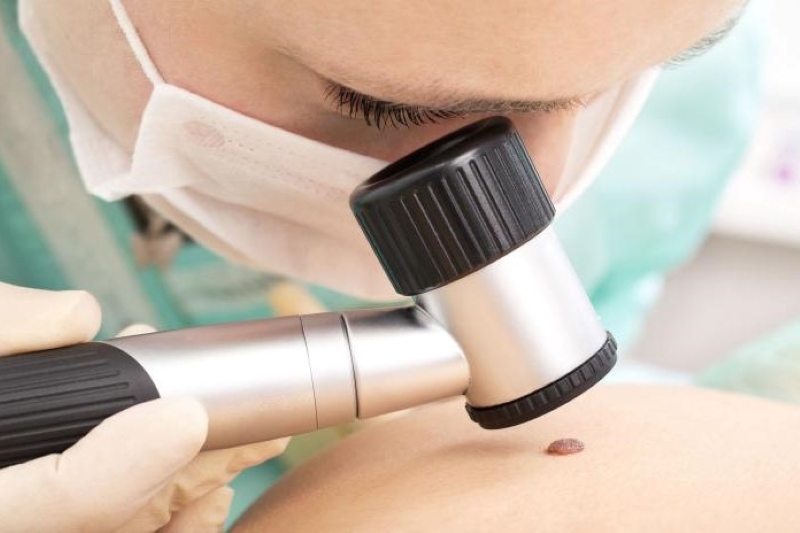
Using a contact microscope (the dermatoscope) connected to a light source, the dermatologist can visualise the anatomical micro-structures of a skin neoformation, located between the epidermis and the dermis.
This direct assessment, in an absolutely non-invasive and painless manner, will make it possible to identify the suspicious features of melanocytic lesions, i.e. correlated to specific histological alterations.
How is epiluminescence performed?
Epiluminescence microscopy can be performed either with a microscope for direct observation of the skin, or using a camera with variable magnification, connected to a monitor for a digitised view of the skin lesion.
The instrument is placed on the skin, after application of a contrast medium (a liquid), and will return the most microscopic features of moles, lesions, keratoses, epitheliomas.
Thanks to dedicated software, it will be possible to carry out a screening of moles or other neoformations and, if the dermatologist deems it appropriate, also the mapping of atypical ones to monitor their evolution over time.
Epiluminescence, if performed correctly, greatly increases the capacity for early detection of melanoma compared to observation alone, as well as reducing the number of unnecessary surgical removals due to its high diagnostic accuracy.
When to do epiluminescence?
The epiluminescence mole test is a check-up indicated by the dermatologist based on the specific characteristics and risk factors of each patient.
In general, it is recommended
- in the presence of a large number of moles
- when a change in the shape, colour or size of a mole is noticed;
- if there is a family history of melanoma;
- in the event of prolonged exposure to the sun or following sunburn;
- when the person has special skin characteristics such as a very fair complexion and is therefore more exposed to the risks of skin cancer.
How to prevent skin cancer?
Periodic mole self-examination is certainly one of the main tools we have to detect a suspicious lesion on the skin at an early stage.
Read Also
Emergency Live Even More…Live: Download The New Free App Of Your Newspaper For IOS And Android
Melanoma: Prevention And Dermatological Examinations Are Essential Against Skin Cancer
Nail Melanoma: Prevention And Early Diagnosis
Dermatological Examination For Checking Moles: When To Do It
What Is A Tumour And How It Forms
Rare Diseases: New Hope For Erdheim-Chester Disease
How To Recognise And Treat Melanoma
Moles: Knowing Them To Recognise Melanoma
Skin Melanoma: Types, Symptoms, Diagnosis And The Latest Treatments
Nevi: What They Are And How To Recognise Melanocytic Moles
Bluish Color Of Baby’s Skin: Could Be Tricuspid Atresia
Skin Diseases: Xeroderma Pigmentosum
Basal Cell Carcinoma, How Can It Be Recognised?
Autoimmune Diseases: Care And Treatment Of Vitiligo
Epidermolysis Bullosa And Skin Cancers: Diagnosis And Treatment
SkinNeutrAll®: Checkmate For Skin-Damaging And Flammable Substances
Healing Wounds And Perfusion Oximeter, New Skin-Like Sensor Can Map Blood-Oxygen Levels
Psoriasis, An Ageless Skin Disease
Psoriasis: It Gets Worse In Winter, But It’s Not Just The Cold That’s To Blame
Childhood Psoriasis: What It Is, What The Symptoms Are And How To Treat It
Topical Treatments For Psoriasis: Recommended Over-The-Counter And Prescription Options
What Are The Different Types Of Psoriasis?
Phototherapy For The Treatment Of Psoriasis: What It Is And When It Is Needed
Skin Diseases: How To Treat Psoriasis?
Skin Cancers: Prevention And Care


