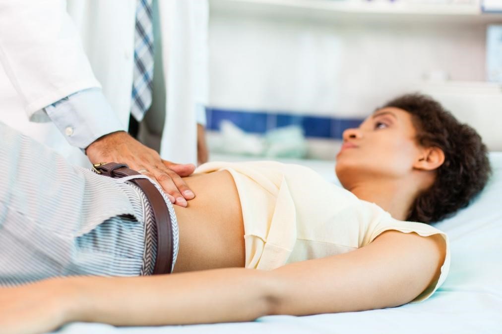
Esophagogastroduodenoscopy (EGD Test): how it is performed
Esophagogastroduodenoscopy is an instrumental test that allows the doctor to look and investigate inside the digestive system to detect any pathologies affecting the oesophagus, stomach and duodenum
Although not recently introduced clinically, in recent years, thanks to technical improvements in instrumentation, it has become increasingly used in the specialist field.
Why and when is oesophagogastroduodenoscopy used
There are several possible indications for the use of oesophagogastroduodenoscopy (EGD Test).
It can be performed in an emergency or election to detect or rule out the presence of disease when the person complains of pain, nausea, vomiting or digestive difficulties.
It is not useful and does not assess changes in the motility of the oesophagus, stomach and duodenum.
In emergencies, EGDS is indicated in the extraction of foreign bodies swallowed voluntarily or involuntarily (e.g. razor blades, coins, ova containing drugs, pen caps, chicken bones and fish bones lodged in the oesophagus) or to stop, by caustication, the bleeding of internal lesions such as ulcers or varices.
In these cases, the test will always be conducted in a hospital environment and with the assistance of an anaesthetist and a supportive nursing team, as these patients are often unprepared (and often not fasting) and in a precarious clinical situation, e.g. due to heavy bleeding.
Oesophagogastroduodenoscopy carried out in elective, i.e. scheduled, outpatient or short-stay (so-called day hospital) settings.
The most frequent indications for which it is useful to undergo this test are:
- in children: repeated vomiting and growth retardation;
- in adults: abdominal pain, burning and hyperacidity, vomiting, slimming and anaemia for which there is no other clinical explanation, periodic monitoring of oesophagogastric varices in cirrhotic patients, monitoring of potentially cancerous lesions, monitoring after surgical or endoscopic removal of lesions or parts of the stomach and oesophagus, placement of oesophageal or gastric prostheses in the course of malignant diseases.
During endoscopy carried out in elective cases, it is also possible to take small tissue samples (biopsies) for the histological diagnosis of suspicious lesions (ulcers, polyps, Barrett’s oesophagus, etc.) but also for the typing of gastritis and the search for Helicobacter pylori, a germ that often causes ulcers and gastritis and their recurrence after treatment.
Although it is an easy test to perform and low risk for the patient, it is preferable for the indication to perform it to be given by medical specialists or in any case to be discussed with the endoscopist, also to better direct the latter on what and where to look for, as well as to minimise the performance of ‘useless’ tests.
How oesophagogastroduodenoscopy is performed
Oesophagogastroduodenoscopy is a test that uses a thin flexible tube (just over a metre long and about one centimetre in calibre) that is introduced through the mouth and slowly moved down the various segments of the digestive tract.
The small tube, which is extremely flexible especially at the tip, is guided by the operator from the outside using a few controls and is connected to a halogen light source that illuminates the inside of the various tracts to be explored.
Thanks to the optical fibres it contains, the operator is able to see through an eyepiece or, more recently, directly on a screen, each part of the viscera being explored. Moreover, inside the tube, between the optic fibres, pass some thin channels through which the operator can introduce a wide range of instruments, such as forceps for biopsies, needles to causticate bleeding lesions, forceps to grasp swallowed objects; it is also possible to introduce water to wash the walls of the viscera, air to dilate them, or even to suck in excess liquids that, for example, obstruct vision.
As a general rule (there may be variations depending on the centre), the patient must lie on his or her left side; local anaesthesia of the pharynx is administered using sprays or pills to dissolve in the mouth, so as to reduce the brief discomfort of passing the instrument through the throat.
Usually a small drip is applied into a vein in the arm, which can be used to administer sedatives or other drugs as appropriate.
The small tube is introduced into the mouth through a disposable mouthpiece, which the patient clasps between his or her teeth to allow easier sliding and also to avoid unintentional biting the expensive and delicate equipment.
It is usually preferred not to put the patient completely to sleep because some slight cooperation may be required during the test (such as holding air, changing position on the couch, etc.).
A diagnostic (i.e. routine) test takes only a few minutes; it may take slightly longer in the event of special difficulties, such as patient intolerance, the need to wash the stomach soiled with food residues or aspirate excess fluid from the viscera, biopsy samples or other operative manoeuvres.
How to prepare for oesophagogastroduodenoscopy
Most endoscopy centres require patients, at the time of booking, to sign an informed consent form (required by law from every patient undergoing so-called ‘invasive’ medical procedures), to undergo a pre-examination or in any case to present certain investigations (e.g. electrocardiogram, routine laboratory investigations, hepatitis virus detection, etc.) at the time of the test.
The test in elective, i.e. scheduled, should be carried out with the patient fasting from the evening before.
Only a few sips of water are allowed on the morning of the test and, if possible, no pills and no smoking.
If sedation is carried out, it is not advisable to drive for about two hours, and one day’s rest and fasting is required if polyps have been removed or bloody manoeuvres performed.
Usually the patient does not experience any discomfort after the test, apart from occasional transient discomfort when swallowing or, rarely, a slight swelling of the salivary glands, which also resolves quickly.
Risks of oesophagogastroduodenoscopy
Serious complications from the test, such as rupture of the oesophagus or stomach, are now very rare.
A cooperative patient, an experienced endoscopist with a proven team, and good equipment are all factors that help minimise the possibility of complications and failures during the test.
Read Also
Emergency Live Even More…Live: Download The New Free App Of Your Newspaper For IOS And Android
Gastroscopy: What The Examination Is For And How It Is Performed
Gastro-Oesophageal Reflux: Symptoms, Diagnosis And Treatment
Endoscopic Polypectomy: What It Is, When It Is Performed
Straight Leg Raise: The New Manoeuvre To Diagnose Gastro-Oesophageal Reflux Disease
Gastroenterology: Endoscopic Treatment For Gastro-Oesophageal Reflux
Oesophagitis: Symptoms, Diagnosis And Treatment
Gastro-Oesophageal Reflux: Causes And Remedies
Gastroscopy: What It Is And What It Is For


