First aid: initial and hospital treatment of drowning victims
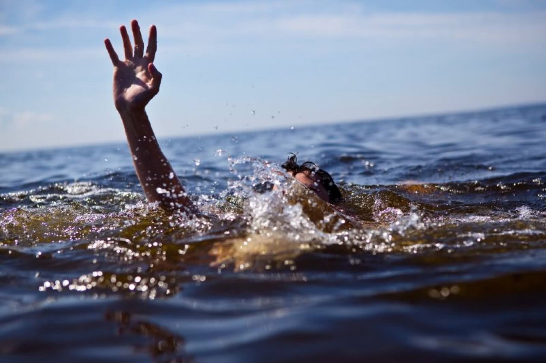
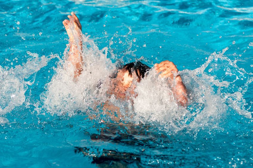
Drowning’ or ‘drowning syndrome’ in medicine refers to a form of acute asphyxia from an external mechanical cause caused by the occupation of the pulmonary alveolar space by water or other liquid introduced through the upper airways, which are completely submerged in such liquid
If the asphyxia is prolonged for a long time, usually several minutes, ‘death by drowning’ occurs, i.e. death due to suffocation by immersion, generally linked to acute hypoxia and acute failure of the right ventricle of the heart.
In some non-fatal cases, drowning can be successfully treated with specific resuscitation manoeuvres
IMPORTANT: If a loved one has been the victim of drowning and you have no idea what to do, first contact emergency services immediately by calling the Single Emergency Number.
Initial treatment of drowning victims
Emergency manoeuvres must be practised and help must be activated as soon as possible by calling Emergency Number.
In the meantime, the rescuer must carefully clear the subject’s airway and, in the absence of spontaneous respiratory activity, begin mouth-to-mouth resuscitation until the patient regains independent breathing.
The search for a heartbeat should be performed after the patient has been returned to shore or lifted onto a float large enough to accommodate both victim and rescuer.
Chest compression manoeuvres performed in water are not effective enough to restore flow.
If the accident occurred in cold water, it is advisable to spend a few extra seconds looking for peripheral pulsations, in order to rule out the presence of marked bradycardia or particularly weak cardiac activity.
A hastily performed cardiac massage can induce ventricular fibrillation and, in fact, worsen cerebral perfusion.
The Heimlich manoeuvre should not be performed unless an airway obstruction caused by some object coexists: drowning victims may swallow considerable amounts of water and the HeimIich manoeuvre may cause them to vomit, with subsequent aspiration, which may worsen the situation.
The head and neck should not be mobilised, particularly if the person drowned after diving into shallow water.
If an injury to the spinal column is suspected, it is necessary to immobilise the patient before transport to avoid possible further damage, in some cases irreversible and disabling, such as that leading to paralysis.
As soon as possible, the patient should be transported to hospital.
Hospital treatment of drowning victims
The hospital staff must prepare the necessary equipment for intubation (laryngoscope, various scalpels, cannulas of various calibre, flexible specils, Magill forceps, syringes to check the patency of the sleeves and to inflate them, aspirator, plaster to fix the endotracheal cannula, appropriate ventilator of the ‘balloon-valve-mask’).
An arterial haemogasanalysis kit and appropriate clothing must be available to ensure the necessary hygienic precautions.
The treatment of drowning victims is based on a rapid initial clinical examination and subsequent classification of the severity of the patient’s condition.
Drowning, the following scheme refers to the post-drowning neurological classification of Modell and Conn:
A) Category A. Awake
- Awake, conscious and oriented patient
B) Category B. Dulling
- Dulling of consciousness, patient is lethargic but can be awakened, purposeful response to painful stimuli
- Patient cannot be awakened, responding abnormally to painful stimuli
C) Category C. Comatose
- C1 Decerebrate-type flexion to painful stimuli
- C2 Decerebrate-type extension to painful stimuli
- C3 Flaccid or absent response to painful stimuli
Drowning, let us now look at the different categories individually
Category A (Awake)
These patients are in an alert state and have a Glasgow Coma Scale (GCS) of 14, indicative of minimal hypoxic damage.
Although the victims in this category are basically healthy, they must still be hospitalised and placed under continuous observation for 12-24 hours to allow early intervention in the event of sudden deterioration of pulmonary or neurological function, a deterioration that must always be anticipated even in the case of an apparently completely healthy subject.
The examinations must include:
- a complete blood count,
- a determination of serum electrolytes and blood glucose,
- a chest X-ray,
- an arterial blood gas analysis,
- sputum culture tests,
- determination of coagulation times.
Drug-toxicological screening may also be necessary.
In the case of suspected neck trauma, an X-ray and/or CT scan of the spine should be performed.
In the case of head trauma or fractures, imaging must obviously also investigate the skull and fractures.
The treatment of patients who fall into this category is basically symptomatic.
Oxygen can be administered, via cannula or mask, in order to maintain a PaO2 above 60 mmHg.
Spirometry may be useful.
The possible aspiration of foreign bodies can be confirmed by a chest X-ray or endoscopy.
Bronchospasm can be treated with β2-adrenergic drugs by aerosol.
Lastly, it is important to ensure venous access, which allows the hydro-electrolyte balance to be controlled and rapid intervention in the event of a deterioration in the clinical condition.
A worsening neurological condition may depend on many factors, such as:
- hypoxemia, secondary to deterioration of pulmonary function ;
- increased intracranial pressure (ICP), secondary to hypoxia;
- medication or drug intake prior to the accident;
- previous metabolic, respiratory, coagulative and/or cardiological diseases.
If the clinical condition remains stable and there is no worsening of neurological or pulmonary function within 12-24 hours, the patient can generally be discharged, except in rare cases.
A medical check-up within 2-3 days is strongly recommended.
Category B (Drowsiness)
These patients are in a state of dullness, or semi-consciousness, but can be awakened.
The GCS score is usually between 10 and 13, indicative of a more severe and prolonged episode of asphyxia.
They respond to painful stimuli with purposeful movements, respiratory activity and pupillary reflexes are normal.
They may be irritable and aggressive.
After resuscitation and initial assessment in the emergency department, these patients should be admitted to an intensive care unit (ICU), carefully monitoring the appearance of any alterations in neurological, pulmonary and/or cardiovascular function.
Their hospital stay is generally longer than for category A patients.
All diagnostic tests should be performed and all therapies discussed above in the section on category A patients.
A daily culture of blood, sputum and, if possible, urine samples should be performed.
The administration of vitamin K can improve clotting times.
Antibiotic therapy should only be administered in the presence of positive culture tests for pathogenic bacterial flora.
The patient’s neurological condition can also change rapidly, and the normal routine for patients with head injuries must be observed.
The appearance of pulmonary oedema or intractable metabolic acidosis, and the need to prolong resuscitation manoeuvres (except for patients extracted from very cold water) are usually indicative of severe hypoxia.
Hypoxemia may become refractory to increasing oxygen concentrations in the inspired air.
In order to maintain a PaO2 above 60 mmHg, continuous positive pressure ventilation (CPAP) using a mask or mechanical equipment may be necessary.
It is sometimes necessary to reduce fluid intake, but the plasma osmolality must not exceed 320 mOsm/litre.
Category C (Coma)
The neurological condition of these extremely critical patients is such that they cannot be awakened.
The GCS score is less than 7.
Treatment must basically be directed at maintaining normal oxygenation, ventilation, perfusion, blood pressure, glycaemia and serum electrolytes.
Small animal studies on cerebral resuscitation have raised new hopes for the recovery of comatose patients who have suffered a severe anoxic insult.
The aim of cerebral resuscitation manoeuvres is to prevent an increase in ICP and to preserve vital but non-functional neurons.
Treatment might include hypothermia, hyperventilation, calcium channel blockers, barbiturates, muscle relaxation or paralysis, etomidate, fluorocarbon infusion.
Unfortunately, the results of cerebral resuscitation manoeuvres are patchy, and it is still controversial which therapy is preferred.
A serious ethical problem relates to the doubt that cerebral resuscitation does not improve the quality of life of patients, but merely delays their death by increasing the number of people in a persistent vegetative state.
The following paragraphs are based on Conn’s recommendations on brain resuscitation.
In this context the prefix ‘HYPER’ is used non-randomly, as patients with severe brain injuries are frequently
- hyperhydrated,
- hyperpyretic,
- hyperexcitable,
- hyperrigid,
- hyperventilated.
Hyperhydration
Hyperhydration can contribute to an increase in ICP and the onset of pulmonary oedema.
In an attempt to prevent this, diuretics are usually administered.
Haemodynamic monitoring is performed to avoid excessive fluid restriction, which could induce renal failure.
Small doses of dopamine (less than 5 μg/kg/min) stimulate renal dopamine receptors, increasing renal perfusion and may thus stimulate urine formation.
Diuresis, however, should not be forced until the serum osmolarity exceeds 320 mOsm/litre.
Performing invasive haemodynamic monitoring requires the insertion of a pulmonary artery catheter, which allows central venous pressure, pulmonary artery pressure and pulmonary wedge pressure to be recorded.
If the arterial pressure is unstable, or if numerous ABGs are performed, the insertion of an arterial catheter may also be necessary.
In the 1980s, ICP was widely practised in order to prevent or control the onset of intracranial hypertension.
Currently, this procedure is most frequently applied in patients who fall into categories A and B and show signs of mental and neurological deterioration.
It is hoped that hyperventilation and the use of osmotic diuretics and thiopental can regress cerebral oedema secondary to ischaemia.
Unfortunately, even effective control of ICP does not guarantee survival without sequelae.
Hyperventilation
Patients requiring mechanical ventilation should be hyperventilated, keeping the paC02 between 25 and 30 mmHg.
Cerebral vascular resistance is controlled by arteriolar tone, which is modified by changes in pH.
Since pH is influenced by PaCO2 values, hyperventilation induces vasoconstriction and reduces ICP values.
The tidal volume can be set from 10 to 15 ml/kg, at the ventilatory rate required to induce the desired PaCO2 reduction.
Tissue oxygenation is an important goal in the treatment of patients with more severe lung impairment.
It would be optimal, but not always possible, to maintain arterial oxygen saturation (SaO2) around 96% (PaO2 of 100 mmHg).
The use of positive end-expiratory pressure (PEEP) is a useful means of ensuring adequate oxygenation (PaO2 above 60 mmHg).
In adults and older children, PEEP values should be increased by 5 cm H2O at a time until adequate oxygenation is achieved.
In younger patients, subsequent increases should be smaller.
Hyperpyrexia
The induction of hypothermia (body temperature of 30± 1°C or lower) has been proposed for brain-injured and comatose patients because it can reduce the metabolic demands of the brain and ICP.
Hypothermia, induced before cerebral ischaemia, is known to exert a protective effect on the brain.
Despite this, this procedure did not improve the neurological condition of patients who had already undergone cerebral hypoxia and may, on the contrary, induce complications, such as suppression of the normal immune response, a leftward shift in the haemoglobin dissociation curve, and cardiac arrhythmias.
If the body temperature is high, normothermia must be restored, with the administration of antipyretics and the use of cooling mattresses, as fever induces an increase in oxygen consumption.
Hyper-excitability
Barbiturates are believed to reduce ICP by inducing vasoconstriction, suppressing convulsive activity, and slowing down cerebral metabolism.
Thiopental is probably the only barbiturate capable of removing oxygen free radicals.
The induction of a pharmacological coma with barbiturates has not been shown to improve survival or the evolution of neurological conditions in drowning victims with severe brain damage and may, on the contrary, accentuate cardiovascular instability.
For these reasons, the administration of barbiturates is no longer part of the recommended treatment; instead, these drugs are used to control convulsive seizures.
The administration of steroids has been proposed, in cases of failed drowning, in the hope of reducing ICP, but subsequent studies have shown them to be ineffective.
In addition, these drugs may interfere with the immune response to bacterial infections, leading to a higher incidence of sepsis.
Hyperrigidity
Decerebrate and decorticated postural rigidity is a sign of intracranial hypertension.
Increased ICP may be secondary to cerebral oedema from hypoxia, mechanical ventilation and PEEP, coughing, Trendelemburg position.
Aspiration manoeuvres may lead to an increase in ICP for up to 30 minutes.
ICP can be reduced in patients requiring mechanical ventilation by administration of sedatives and paralysing agents.
Read Also:
Emergency Live Even More…Live: Download The New Free App Of Your Newspaper For IOS And Android
Drowning Resuscitation For Surfers
ERC 2018 – Nefeli Saves Lives In Greece
First Aid In Drowning Children, New Intervention Modality Suggestion
Water Rescue Dogs: How Are They Trained?
Drowning Prevention And Water Rescue: The Rip Current
RLSS UK Deploys Innovative Technologies And The Use Of Drones To Support Water Rescues / VIDEO


