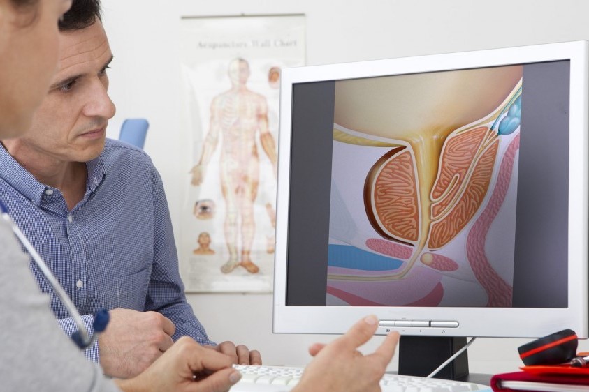
Fusion prostate biopsy: how the examination is performed
Prostate biopsy using the Fusion technique is a method that allows targeted diagnosis of prostate cancer: the most frequent neoplasm in men, with an estimated 36,000 new diagnoses in Italy based on the latest data from 2020
Neoplasms which, however, since 2015 have recorded a 14.6% reduction in the mortality rate, also in view of the new, increasingly advanced diagnostic techniques including, precisely, fusion biopsy.
What is Fusion prostate biopsy
Fusion prostate biopsy is a biopsy procedure, i.e. collecting specimens for diagnostic purposes, in which images of the prostate obtained during a transrectal ultrasound scan are ‘fused’ using special software with those of a Multiparametric Magnetic Resonance Imaging (MRI) scan of the prostate previously performed by the patient.
Fusion prostate biopsy, how the examination is performed
The examination lasts about 30 minutes.
After having the patient lie sideways on the couch, the doctor performs a transrectal ultrasound scan.
On the ultrasound monitor, the 3D images collected in real time by the probe are merged with those of the MRI scan previously taken by the patient and loaded into the ultrasound machine.
At this point, the doctor proceeds under local anaesthesia and samples the suspicious areas with the probe.
The area to be sampled is identified on the ultrasound monitor as a target to be targeted.
How to prepare for the examination
In order to be able to carry out the procedure, it is necessary for the patient to follow certain steps prescribed by the doctor beforehand.
Carry out and bring with you for examination:
- MRI of the prostate;
- blood tests including total PSA, free PSA, complete blood count, prothrombin time (PT), partial thromboplastin time (PTT), INR;
- urine tests and urine culture;
- undertake antibiotic prophylaxis in order to avoid infection following the examination;
- discontinue any anti-aggregant or anti-coagulant therapy or follow a revisit therapy, in order to minimise the risk of procedure-related bleeding;
- undergo an enema to clean the rectal canal through which the ultrasound probe will pass;
- follow a 6-hour fast (this is only if the examination is carried out under sedation).
The advantages of the fusion prostate biopsy technique
The important advantages of performing this procedure are
- high sensitivity and more accurate diagnosis: by integrating and enhancing ultrasound images with those of MRI, fusion biopsy has, compared to the traditional ultrasound procedure, a higher sensitivity and precision in the diagnosis of the most aggressive prostate tumours;
- 3D mapping of the biopsies which, if neoplasms are detected, allows an approximate reconstruction of their volume, position and characteristics, so that the most suitable treatment plan can also be drawn up on the basis of this
- less risk of complications: reducing the number of samples also reduces the consequent risk of urinary infections, prostate inflammation (prostatitis) and the presence of blood in urine (haematuria), semen (haemospermia) or from the rectum (rectorrhagia);
- less sampling and more precise: fusion biopsy allows sampling only in areas identified as suspicious, as opposed to traditional transrectal ultrasound (TRUS) which involves ‘blind’ sampling at 12 to 18 points of the prostate, with the high risk of missing or partially identifying neoplastic areas.
Screening and prevention
According to the report ‘The Numbers of Cancer 2021’, the fact that prostate cancer has become the most frequent type of cancer in the Western male population is linked to the increased likelihood of diagnosis through early screening.
In fact, according to the report, prostate cancer is the cancer disease with the highest prevalence in men (564,000 cases).
For many men, unfortunately, the admonition to overcome embarrassment and visit the urologist still applies.
The doctor, however, is often already able to detect an increase in the volume and consistency of the prostate gland by means of an objective examination, which may be further investigated with
- blood tests (PSA);
- multi-parametric magnetic resonance imaging of the prostate;
- biopsies.
Multiparametric prostate MRI (MRI)
In particular, multi-parametric prostate MRI (MRImp) is a very sensitive examination for prostate cancer which, as the expression itself indicates, acquires multiple morphological, functional and metabolic parameters, making it the best non-invasive investigation to identify prostate neoplasms which, in any case, must be confirmed by biopsy.
Read Also:
Emergency Live Even More…Live: Download The New Free App Of Your Newspaper For IOS And Android
Prostatitis: Symptoms, Causes And Diagnosis
Colour Changes In The Urine: When To Consult A Doctor
Acute Hepatitis And Kidney Injury Due To Energy Drink Consuption: Case Report
Bladder Cancer: Symptoms And Risk Factors
Enlarged Prostate: From Diagnosis To Treatment
Male Pathologies: What Is Varicocele And How To Treat It
Continence Care In UK: NHS Guidelines For Best Practice
The Symptoms, Diagnosis And Treatment Of Bladder Cancer


