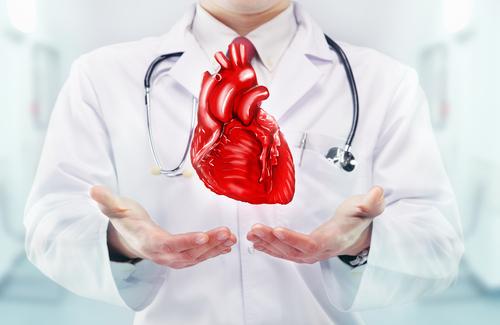
Inflammations of the heart: myocarditis, infective endocarditis and pericarditis
Let’s talk about inflammation of the heart: the heart, the nucleus of the circulatory system, begins to beat around 16 days after conception, and from that moment on its continuous motion of contraction and release accompanies us for the rest of our lives
It receives venous blood from the periphery, feeds it into the pulmonary circulation to oxygenate it, and then pumps oxygen-rich blood into the aorta and arteries to carry it to the body’s organs and tissues.
Every minute, the heart beats an average of 60 to 100 times and can carry as much as 5 to 6 litres of blood.
Anatomy of the heart
The heart, which is located in the chest between the two lungs, is about the size of a closed fist and weighs about 200-300 grams.
Its structure consists of three layers:
- Pericardium: this is the thin surface membrane that covers it externally and which also envelops the large incoming and outgoing blood vessels;
- Myocardium: the muscular tissue that makes up the walls of the heart;
- Endocardium: is the thin lining of the inner walls of the heart cavities and valves.
The heart has four distinct chambers, two atria (right and left) and two ventricles (right and left).
Separating the two atria and the two ventricles are the interatrial and interventricular septum, respectively.
The right atrium and its corresponding ventricle are responsible for receiving oxygen-poor, carbon dioxide-rich venous blood and pumping it into the lungs, while the left atrium and ventricle are responsible for pumping oxygenated blood first into the aorta and then into the arteries, ready for distribution throughout the body.
Four valves are responsible for regulating blood flow within the heart:
- tricuspid: between the atrium and right ventricle
- mitral valve: between the atrium and left ventricle
- pulmonary: between the right ventricle and the pulmonary artery
- aortic: between the left ventricle and aorta
Valves open and close according to changes in blood pressure produced by the relaxation and contraction of the myocardium and prevent blood from flowing back in the wrong direction.
Inflammations of the heart
Myocarditis, pericarditis and endocarditis are the inflammations or infections that can affect the myocardium, pericardium and endocardium, respectively.
Inflammations of the heart: myocarditis
What is myocarditis?
Myocarditis is an inflammation of the heart muscle. It occurs mostly as a result of viral infections, but also following exposure to drugs or other toxic substances (e.g. certain chemotherapeutic agents) or due to autoimmune diseases.
Myocarditis can present itself in very variable ways and, likewise, can have very different evolutions: complete recovery is possible or, sometimes, cardiac function may be compromised.
In forms associated with viral infections, myocarditis is caused by two possible mechanisms: the direct action of the infectious agent, which damages and destroys muscle cells, but also the intervention of immune cells.
Myocarditis may be associated with pericarditis if the inflammation also involves the pericardium.
Inflammations of the heart: what are the causes of myocarditis?
The main conditions from which myocarditis can develop are:
- Viral infections (such as Coxsackievirus, Cytomegalovirus, Hepatitis C virus, Herpes virus, HIV, Adenovirus, Parvovirus…) that cause damage to myocardial cells either by a direct mechanism or by activation of the immune system.
- More rarely bacterial, fungal and protozoal infections.
- Exposure to drugs and toxic substances: these can cause direct damage to myocardial cells (e.g. cocaine and amphetamines) or allergic reactions and activation of the immune system (drugs including certain chemotherapeutic drugs, antibiotics or antipsychotics).
- Autoimmune and inflammatory diseases (e.g. systemic lupus erythematosus, rheumatoid arthritis, scleroderma, sarcoidosis).
What are the symptoms of myocarditis?
The manifestations of myocarditis can be very diverse. The most frequent symptom is chest pain, similar to that of a heart attack.
Other frequent symptoms are shortness of breath, fever, fainting and loss of consciousness.
Flu-like symptoms, sore throat and other respiratory tract infections or gastrointestinal disorders may have occurred in the preceding days and weeks.
In complicated forms there may be malignant arrhythmias and signs and symptoms of severe cardiac dysfunction.
Diagnosis of myocarditis: what tests for this cardiac inflammation?
When the history and symptoms suggest a possible myocarditis, the tests that allow the diagnosis are:
- Electrocardiogram (ECG);
- Blood tests, in particular cardiac enzymes and inflammatory markers;
- Echocardiogram: allows the contractile function of the heart to be assessed;
- In stable patients, the examination that allows a non-invasive diagnosis of myocarditis is cardiac magnetic resonance imaging: in addition to assessing the contractile function of the heart, it allows areas of inflammation of the myocardium and the presence of any scars to be visualised; it is also useful in subsequent months to assess the recovery and evolution of the myocarditis;
- In unstable patients, with complicated forms, or if specific causes are suspected, an endomyocardial biopsy, a sampling of a small portion of heart muscle for laboratory analysis, may be indicated.
- In some patients, coronary arteryography or CT angiography of the coronary arteries may be necessary to exclude significant coronary artery disease.
Inflammations of the heart: How is myocarditis treated?
Hospitalization for initial monitoring and administration of therapy is generally indicated.
In most cases, the therapy is standard heart failure therapy.
In complicated forms, admission to intensive care is required, and in addition to drug therapy, mechanical systems may be needed to support the circulatory system or treat arrhythmias.
If a specific cause is found, targeted treatment or immunosuppressive therapy may be indicated.
Patients suffering from myocarditis are advised to abstain from physical activity for at least 3-6 months, and in any case until normalisation of subsequent investigations and blood tests.
Can myocarditis be prevented?
Unfortunately, there are no real measures that can be taken to prevent the onset of myocarditis.
Inflammations of the heart: pericarditis
What is pericarditis?
Pericarditis is an inflammation affecting the pericardium, the membrane lining the heart and the origin of the great vessels.
The pericardium consists of two sheets, between which is a thin layer of fluid, the pericardial fluid.
Inflammation may or may not result in an increase in fluid between the two membranes (in this case we speak of pericardial effusion).
If the pericardial effusion is abundant and its formation is sudden, it can impede the filling of the heart cavities.
This is known as cardiac tamponade, a condition that requires prompt intervention to drain the excess pericardial fluid.
In rare cases, as a result of inflammation, the pericardium thickens and stiffens, leading to constrictive pericarditis, which prevents proper expansion of the heart.
This is not an emergency situation in this case, but still requires rapid evaluation by a specialist.
After a first episode of acute pericarditis, in some cases it is possible that a second episode, or relapse, may occur, which is very similar to the first.
What are the causes of pericarditis?
There can be several triggering factors behind pericarditis:
- Infectious causes: viruses (common); bacteria (mainly mycobacteria from tuberculosis, other bacterial agents are rare); rarely fungi and other pathogens.
- Non-infectious causes: tumours, advanced kidney failure or autoimmune diseases (e.g. systemic lupus erythematosus etc); drugs (including antibiotics and antineoplastics); radiation treatment; trauma or injury (also related to diagnostic or therapeutic procedures involving the pericardium.
What are the symptoms of pericarditis?
The most characteristic symptom of pericarditis is chest pain. It is a pain with absolutely peculiar characteristics: more intense in the supine position and relieved by sitting and reclining forward; it varies with breathing and coughing.
Other symptoms may be related to those of the underlying cause.
Diagnosis of pericarditis: what tests should be done?
The following tests are necessary to make a diagnosis of pericarditis:
- Electrocardiogram (ECG): changes in cardiac electrical activity are present in more than half of all cases of pericarditis
- Chest X-ray
- Blood tests: mainly elevation of inflammatory indices
- Transthoracic echocardiogram: this can suggest inflammation of the pericardium if it is more ‘reflective’ and also allows the presence of pericardial effusion to be detected and quantified.
How is pericarditis treated?
If the symptoms suggest a specific cause, this should be investigated and treated appropriately.
In all other cases, it is not necessary to investigate the cause and treatment with non-steroidal anti-inflammatory drugs (NSAIDs), in particular acetylsalicylic acid or ibuprofen, is given for several weeks, with the dose being progressively reduced.
Colchicine is combined to reduce the risk of recurrence. Symptoms usually subside within a few days.
If NSAIDs are ineffective or contraindicated, corticosteroids are prescribed. In general, corticosteroids represent a second line of treatment because they are associated with the risk of chronic evolution.
For patients requiring long-term therapy with high doses of corticosteroids, the use of other therapies (azathioprine, anakinra and intravenous immunoglobulins) may be considered.
Can pericarditis be prevented?
As in the case of myocarditis, there are no measures that can be taken to prevent pericarditis.
Inflammations of the heart: Infective endocarditis
What is infective endocarditis?
Endocarditis is an inflammation of the endocardium.
We focus on the infectious form, but remember that there is also non-infectious endocarditis (due to inflammatory or autoimmune diseases or pathologies, such as neoplasms or immune deficiencies, that promote thrombotic deposits).
Endocarditis most often affects the heart valves, but can also occur at shunts or other abnormal communications between cardiac cavities.
This pathology can alter the structure and function of the valves, which can lead to a haemodynamic overload of the heart cavities.
It can also cause embolisation (due to the detachment of infected material) and vascular damage outside the heart.
What are the causes of infective endocarditis?
The characteristic lesion of infective endocarditis is “vegetation”, i.e. a deposit of fibrinous material and platelets attached to the endocardium, in which the microorganisms that cause endocarditis nest and multiply.
The microorganisms that cause infective endocarditis are bacteria and fungi that enter the bloodstream via the mouth, skin, urine or intestines and reach the heart.
The most frequent forms of infective endocarditis are bacterial.
Those at highest risk of developing infective endocarditis are:
- Patients who have already had infective endocarditis;
- Patients with prosthetic valves or other prosthetic material;
- Patients with certain types of congenital heart disease, or those in which uncorrected alterations remain.
Other characteristics that increase the risk of contracting endocarditis are: other forms of valve disease, intravenous drug use or the presence of haemodialysis catheters or other central venous accesses.
What are the symptoms of infective endocarditis?
The infection may develop more suddenly and aggressively or more gradually and subtly.
The signs and symptoms of endocarditis are related to the systemic infectious state and activation of the immune system, the growth of vegetations that damage or prevent the proper functioning of the heart valves, and finally the possible detachment of fragments of vegetation that reach other organs (septic embolisms).
In general, one can distinguish
- symptoms of the infectious state: fever, headache, asthenia, malaise, lack of appetite and weight loss, nausea and vomiting, bone and muscle pain;
- symptoms and signs related to the involvement of cardiac structures, including: difficulty breathing, swelling of the ankles and legs, less frequently chest pain; onset of a new heart murmur;
- symptoms and signs resulting from septic embolisation or immunological phenomena: abdominal and joint pain, headaches, back pain, stroke and other neurological changes; small skin haemorrhages, painful skin nodules, peripheral ischaemia and several others, nowadays very rare.
Diagnosis of infective endocarditis: what tests should be done?
Making a diagnosis of infective endocarditis can be a difficult and complex process, requiring a great deal of clinical attention and analytical skills on the part of doctors.
An initial diagnostic suspicion may emerge if auscultation of the heart of a patient with fever detects a new-onset murmur.
Such a murmur is caused by turbulence in the blood flow, which may be the result of valve malfunction.
If there is a clinical suspicion, the doctor may then prescribe further investigations to establish the diagnosis.
Blood tests may be prescribed to detect changes compatible with endocarditis, in particular:
- bacteria or other microorganisms are sought in the blood, using blood cultures;
- an increase in inflammatory indices.
For the diagnosis of endocarditis, the echocardiogram plays a fundamental role.
This is an examination that uses ultrasound to examine the cardiac cavities and valves, and above all allows direct visualisation of the endocardial vegetations.
Initially, a transthoracic echocardiogram is performed.
Subsequently, a transesophageal echocardiogram may also be requested.
In this case, the ultrasound probe is introduced from the mouth into the oesophagus, allowing better visualisation of the cardiac structures.
This allows the following to be assessed
- Possible valvular lesions;
- Characteristics of the vegetations (size and morphology) and the consequent risk of embolisation;
- Possible complications, such as the formation of aneurysms, pseudoaneurysms, fistulas or abscesses.
Other tests that may be prescribed include:
- electrocardiogram (ECG);
- chest X-ray;
- CT scan with or without contrast medium, PET scan, nuclear magnetic resonance; these are useful in improving the diagnostic picture, as they allow the detection of any extracardiac septic localisation, or cardiac and vascular complications; PET scan can also play a fundamental role in the diagnosis of endocarditis in the presence of valve prostheses, pacemakers and defibrillators.
How is infective endocarditis treated?
The treatment of infective endocarditis is extremely complex and requires in-depth expertise, which is why it must be based on a multidisciplinary approach, with a team of different specialists working together to devise the most appropriate course of treatment.
The treatment, which lasts several weeks, involves targeted antibiotic therapy to combat the infectious agent isolated from blood cultures.
In the event of negative blood cultures, empirical antibiotic therapy is carried out, i.e. using an antibiotic with a broad spectrum of action or one that acts against the presumed infectious agent.
In the presence of signs of heart failure, vegetations with a high embolic risk or in case of insufficient control of the infectious state, surgery is resorted to: surgery is aimed at replacing valves and repairing the damage done by any complications.
Can infective endocarditis be prevented?
The main preventive measures are aimed at minimising, ideally avoiding, bacteremia and the subsequent localisation of bacteria in the endothelium, particularly for the high- and intermediate-risk patient categories outlined above.
They include:
Special attention to oral hygiene, with regular dental visits;
- Antibiotic treatment of any bacterial infections, always under medical supervision and avoiding self-medication, which can promote the emergence of bacterial resistance without eradicating the infection;
- Careful attention to skin hygiene and thorough disinfection of wounds;
- avoid piercings and tattoos.
Antibiotic prophylaxis of endocarditis is only recommended in high-risk categories of patients, before performing dental procedures that require manipulation of gum tissue or perforation of the oral mucosa.
Read Also:
Study In European Heart Journal: Drones Faster Than Ambulances At Delivering Defibrillators
Arrhythmias, When The Heart ‘Stutters’: Extrasystoles


