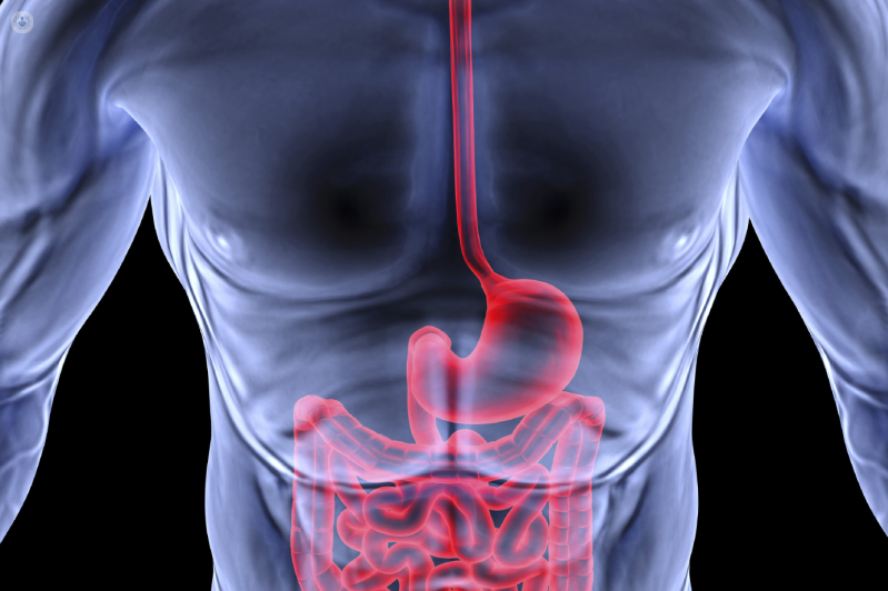
Intestinal ischaemia: survival, tests, treatment, aftercare
In medicine, ‘intestinal ischaemia’ refers to an alteration of blood circulation in the tissues of the intestine, caused by various causes, such as the occlusion of an artery that brings oxygenated blood to the intestine, but also the alteration of intestinal venous flow
Types of intestinal ischaemia:
A distinction is therefore made between venous or arterial intestinal ischaemia, as well as acute or chronic intestinal ischaemia and also occlusive and non-occlusive intestinal ischaemia.
As a result of the altered circulation, the intestinal mucosa has a reduced supply of nutrients and oxygen, with the result that – if the blood flow is not quickly restored – the intestinal mucosa goes into ‘necrosis’ (i.e. dies), leading to the picture of an ‘intestinal infarction’, a complication that can be lethal.
It should be remembered that the intestinal mucosa has a high demand for blood flow (it receives almost a quarter of the entire cardiac output), which makes it very sensitive to the effects of decreased perfusion: intestinal ischaemia therefore sets in rather quickly and can lead to a series of sequential events, even lethal:
- necrosis of the mucosa
- perforation of the mucosa;
- release of bacteria, toxins and vasoactive mediators;
- myocardial depression;
- systemic inflammatory response syndrome (sepsis and septic shock);
- multi-organ failure;
- patient death.
Necrosis may occur as little as 10 hours after the onset of symptoms.
Mesenteric ischaemia is distinct from ischaemic colitis:
- mesenteric ischaemia: blood flow is altered in the small intestine. Less frequent;
- ischaemic colitis: blood flow is altered in the colon (large intestine). More frequent.
Intestinal ischaemia may occur due to an obstruction or vascular rupture at the level of the three major vessels that vascularise the abdominal organs:
- celiac trunk: irrigates the oesophagus, stomach, proximal duodenum, liver, gallbladder, pancreas and spleen;
- superior mesenteric artery: irrigates the distal duodenum, jejunum, ileum and colon up to the splenic flexure;
- lower mesenteric artery: irrigates the descending colon, sigma and rectum.
Mesenteric blood flow can be impaired at the level of these arteries, but also at the level of the venous vessels that collect blood that is no longer oxygenated, from the intestine.
Causes of acute and chronic intestinal ischaemia, occlusive and non-occlusive
Mesenteric ischaemia may be acute or chronic:
- acute mesenteric ischaemia: the interruption of blood supply is sudden and severe (very little blood reaches the tissue). It is generally more severe;
- chronic mesenteric ischaemia: blood flow to the intestine decreases gradually and progressively. It is generally less serious than acute ischaemia, although what is not a serious condition in an absolute sense.
Acute mesenteric ischaemia has three main causes occurring in the superior mesenteric artery
- occlusion of the artery by a blood clot (embolus) originating in the heart, e.g. in cases of prolonged atrial fibrillation (frequent);
- occlusion of the artery by a thrombus caused by the lesion of an atheroma (cholesterol deposit that narrows arterial vessels suffering from atherosclerosis), e.g. in the case of a spike in blood pressure
- reduction of flow in the artery by abrupt arterial hypotension, which can be induced by shock, heart failure, internal haemorrhage, renal failure, abuse of certain medications or drugs.
The first two situations are called ‘acute occlusive mesenteric ischaemia’, while the third situation is called ‘acute non-occlusive mesenteric ischaemia’.
Chronic mesenteric ischaemia, on the other hand, is almost always caused by an occlusion of the mesenteric artery caused by an atheroma that gradually expands.
In this case, atherosclerosis is therefore the cause of chronic ischaemia: chronic mesenteric ischaemia is therefore always of the ‘non-occlusive’ type.
Intestinal ischaemia from venous causes
Intestinal ischaemia can be caused not only by arterial causes, but also by venous ones: when an obstruction prevents venous blood from leaving the intestine properly, it triggers an accumulation and subsequently a reflux, i.e. the blood ‘flows back’.
The basis of venous obstruction is almost always a blood clot (embolus) that blocks the mesenteric vein or its branches.
Such embolism is generally caused or facilitated by:
- acute or chronic pancreatitis;
- abdominal infection;
- abdominal tumour;
- ulcerative colitis;
- Crohn’s disease;
- diverticulitis;
- abdominal trauma;
- hypercoagulation;
- incorrect anticoagulant therapy (inadequate INR);
- cardiac arrhythmias;
- recent surgery, e.g. after femur fracture.
Intestinal ischaemia from venous causes is also called ‘mesenteric venous thrombosis’
Ischaemia from venous causes is, however, less frequent than arterial ischaemia and, in theory, less severe.
The patients most at risk of mesenteric ischaemia are those with the following characteristics and pathologies:
- men;
- age > 50 years;
- overweight and obesity;
- intestinal obstruction from various causes;
- chronic intestinal constipation;
- faecaloma;
- colon tumours;
- large abdominal tumours;
- megacolon;
- dolichocolon;
- sudden severe arterial hypotension (‘very low blood pressure’);
- arterial embolism;
- coronary artery disease;
- heart failure;
- heart valve disease;
- arterial hypertension;
- atrial fibrillation;
- intestinal volvulus;
- intestinal stricture;
- previous surgery;
- positive history of previous arterial embolism;
- arterial thrombosis (30%);
- generalised atherosclerosis;
- venous thrombosis (15%);
- hypercoagulability;
- pancreatitis;
- diverticulitis;
- chronic inflammation;
- cigarette smoking;
- high-fat diet;
- trauma, especially abdominal trauma (e.g. from road accidents);
- heart failure;
- renal insufficiency;
- portal hypertension;
- decompression sickness;
- heart failure;
- shock;
- cardiopulmonary bypass;
- splanchnic vasoconstriction;
- intestinal adhesions;
- cocaine, amphetamine and methamphetamine use;
- intestinal artery vasculitis;
- systemic lupus erythematosus (SLE);
- sickle-cell anaemia;
- use of: drugs with vasoconstrictor effect, drugs for treating heart disease, drugs for treating migraine, hormonal drugs (such as oestrogen);
- excessive physical exertion, especially prolonged physical exertion.
Symptoms and signs
The first characteristic sign of mesenteric ischaemia is severe pain accompanied by minimal physical findings.
The abdomen remains soft, with little or no pain.
Mild tachycardia may be present.
Later, when necrosis develops, signs of peritonitis appear, with marked abdominal tenderness, defensive reaction, rigidity and absence of bowel sounds.
The faeces may show traces of blood (increasingly likely as the ischaemia progresses), of a different colour depending on the intestinal tract affected: darker brown if the small intestine is affected, gradually more bright red if the lesion affects areas closer to the anus (e.g. descending colon and sigma).
Typical signs of shock develop, which are often followed by death.
The sudden onset of pain suggests an arterial embolism but does not permit its diagnosis, whereas a more gradual onset is typical of venous thrombosis.
Patients with a history of postprandial abdominal complaints (suggesting intestinal angina) may have arterial thrombosis.
Symptoms and signs can be differentiated according to three main factors
- arterial or venous intestinal ischaemia;
- ischaemic colitis or mesenteric ischaemia;
- acute or chronic ischaemia.
Symptoms of ischaemic colitis
When ischaemia affects the descending colon (left colon), there are:
- sudden abdominal pain in the left lower quadrant;
- presence of bright red (if the lower part is affected) or brown (if the upper part is affected) blood in the stool.
When ischaemia affects the ascending colon (right colon) there are:
- sudden right lower quadrant abdominal pain;
- absence of blood in the stool or minimal presence of brown or black blood in the stool.
Symptoms of acute mesenteric ischaemia from arterial causes
When ischaemia acutely affects the small intestine, there are:
- sudden and very intense abdominal pain, especially if the cause is occlusive (e.g. embolus);
- general malaise
- abdominal distension;
- abdominal soreness;
- nausea;
- vomiting;
- abnormal bowel movements;
- urgent need to defecate.
Symptoms of chronic mesenteric ischaemia from arterial causes
When ischaemia affects the small intestine chronically, there are:
- post-prandial abdominal pain (10-30 minutes after meals, peaking after about 2 hours and then gradually decreasing). This pain tends to become more intense over time;
- abdominal cramps;
- drop in body weight (the patient eats less for fear of feeling pain).
Symptoms of mesenteric ischaemia from venous causes
When ischaemia affects the small intestine from venous causes, there are:
- abdominal pain (less intense than with ischaemia from arterial causes)
- general malaise;
- nausea;
- vomiting;
- diarrhoea;
- blood in the stool (not always).
Diagnosis and differential diagnosis of intestinal ischaemia
Early diagnosis is particularly important as mortality increases significantly once intestinal infarction has occurred: early diagnosis generally saves the patient’s life.
Mesenteric ischaemia should be considered in any patient > 50 years of age, with known risk factors or predisposing conditions, who presents with sudden and severe abdominal pain.
Patients with clear peritoneal signs should be sent directly to the operating theatre for both diagnosis and treatment.
In others, selective mesenteric angiography or CT angiography is the diagnostic procedure of choice.
Other imaging studies and serum markers may be altered but are not sensitive and specific in the early stages of the disease, when it is most important to make the diagnosis.
Direct X-ray of the abdomen is useful in the differential diagnosis to exclude other causes of pain (perforated bowel), although in advanced stages of the disease the presence of gas bubbles in the portal vein or intestinal pneumatosis may be observed.
These findings are also visible in CT scans, which can also directly visualise the vascular occlusion more accurately on the venous side.
Echodoppler can sometimes identify an arterial occlusion, but the sensitivity is low. MRI is very accurate in proximal vascular occlusion, but less so in distal vascular occlusion.
Haematochemical examinations
Serum markers (creatine phosphokinase and lactate) increase with necrosis, but are non-specific and late findings.
Neutrophil leucocytosis and occult blood in the faeces are other important parameters for diagnosis. Serious intestinal fatty acid binding protein may perhaps prove useful as an early marker in the future.
Introduction to treatment
If the diagnosis is made during exploratory laparotomy, the options are surgical embolectomy, revascularisation or resection.
A laparotomic ‘second look’ may be necessary to re-evaluate the viability of suspicious areas of the bowel.
If the diagnosis is made by angiography, infusion of the vasodilator papaverine through the angiographic catheter may improve survival in both occlusive and non-occlusive ischaemia.
Papaverine is also useful when surgery is planned and is sometimes also administered during and after surgery.
In addition, thrombolysis or surgical embolectomy may be performed in the event of arterial occlusion.
The appearance of peritoneal signs at any time during evaluation suggests the need for immediate surgical intervention.
Mesenteric venous thrombosis without signs of peritonitis can be treated with papaverine followed by anticoagulants such as heparin and then warfarin.
Patients with arterial embolism or venous thrombosis require long-term anticoagulant therapy with warfarin.
Patients with non-occlusive ischaemia may be treated with anti-platelet therapy.
Specific therapies according to the cause and type of intestinal ischaemia
The specific therapy of intestinal ischaemia varies depending on the cause, severity and type of ischaemia.
Common to all therapies are three objectives
- to restore normal blood flow to the intestine
- to reduce the patient’s painful symptoms;
- surgically remove intestinal tracts that may no longer be viable (necrotic).
Specific therapies for ischaemic colitis
If the cause is atherosclerosis, therapy involves pharmacological treatment:
- anticoagulant;
- vasodilator.
In more severe cases, it may be necessary
- stent angioplasty surgery (the occlusion is removed with a sort of balloon)
- a bypass surgery, to create an ‘alternative route’ that allows blood to still reach the ischaemic tract.
In other cases (not an embolus), the specific cause is intervened on if possible: intestinal volvulus, colon cancer, heart failure, vasculitis, drug abuse… these are all situations that are intervened on to interrupt the ischaemia.
If the damage to the intestine is irreversible, surgery is performed to remove the necrotic intestinal tract.
Specific therapies for acute mesenteric ischaemia from arterial causes
If the cause is an embolus, therapy includes:
- anticoagulant therapy;
- vasodilator therapy;
- embolectomy (if the embolus is not removed with pharmacological remedies).
If the cause is a thrombus, therapy involves angioplasty with a stent.
In other cases (not a blood clot, nor a thrombus), the specific cause is addressed if possible: heart failure, renal failure, occluding tumour, drug abuse… these are all situations in which we intervene to interrupt the ischaemia.
If the damage to the intestine is irreversible, surgery is performed to remove the necrotic intestinal tract.
Specific therapies for chronic mesenteric ischaemia from arterial causes
Therapy includes:
- stent angioplasty surgery (the occlusion is removed with a sort of balloon)
- bypass surgery, to create an ‘alternative route’ that allows blood to still reach the ischaemic tract.
It is important to reduce the atherosclerotic risk (e.g. with diet and statins).
Specific therapies for mesenteric ischaemia from venous causes
Therapy involves taking anticoagulants for 3-6 months (in some cases therapy is for life). In the presence of irreversible damage to the intestine, in addition to anticoagulant therapy, surgery is performed to remove the necrotic intestinal tract.
Postoperative course
The postoperative course basically depends on the patient’s condition, the type of therapy applied and the portion of the intestine that has gone into necrosis. In the case of removal of large parts of the intestine, the hospital stay may be prolonged.
Patients generally return to normal activities within 3-4 weeks, during which they should avoid exertion and follow the diet recommended by their doctor.
Complications of intestinal ischaemia
Intestinal ischaemia, whether it affects the colon or the intestine, whether from occlusive or non-occlusive causes, is a potentially fatal event, especially if acute and especially if diagnosis and treatment are not rapid, leading to intestinal infarction.
In the absence of prompt treatment or if it is very severe, ischaemia can lead to various complications:
- necrosis of the intestinal tract involved (intestinal infarction)
- perforation of the involved intestinal tract;
- intestinal haemorrhage;
- leakage of intestinal contents (digested food or faeces depending on the perforated tract);
- peritonitis (infection of the peritoneum);
- scarring in the affected intestinal tract, with narrowing of the lumen of the intestinal tract that favours future intestinal occlusions;
- myocardial depression;
- systemic inflammatory response syndrome (sepsis and septic shock);
- multi-organ failure;
- patient death from haemorrhage and/or shock and/or sepsis and/or other related causes.
Survival
Survival to acute mesenteric ischaemia is highly variable and strongly influenced by the timeliness of intervention: if diagnosis and treatment take place before the ischaemia leads to intestinal infarction, the prognosis is much better, with a low mortality.
If diagnosis and treatment take place after the intestinal infarction, mortality is generally very high, reaching 70-90%, with variability due to many factors, such as the patient’s age and any other pathologies such as diabetes or coagulopathies: elderly patients with such pathologies have a higher average risk.
Early diagnosis and early treatment, as and more than in other diseases, make the real difference between life and death in this case.
Tips
You can reduce the risk of intestinal ischaemia and recurrences by making a few simple changes to your lifestyle, which help prevent atherosclerosis and other risk factors.
A diet rich in fruit, vegetables and whole grains and reducing the amount of added sugar, carbohydrates, cholesterol and fat is essential.
Fibre should be neither too much nor too little.
It is also recommended to:
- do not smoke;
- lose weight if obese or overweight;
- take regular exercise;
- keep your blood pressure under control;
- avoid abdominal trauma;
- avoid intense exertion;
- avoid binge eating;
- avoid drugs;
- avoid alcohol;
- avoid psycho-physical stress and anger outbursts.
Read Also:
Emergency Live Even More…Live: Download The New Free App Of Your Newspaper For IOS And Android
Peptic Ulcer, Symptoms And Diagnosis
Vomiting Blood: Haemorrhaging Of The Upper Gastrointestinal Tract
Peptic Ulcer, Often Caused By Helicobacter Pylori
Peptic Ulcer: The Differences Between Gastric Ulcer And Duodenal Ulcer
Wales’ Bowel Surgery Death Rate ‘Higher Than Expected’
Irritable Bowel Syndrome (IBS): A Benign Condition To Keep Under Control
Ulcerative Colitis: Is There A Cure?
Colitis And Irritable Bowel Syndrome: What Is The Difference And How To Distinguish Between Them?
Irritable Bowel Syndrome: The Symptoms It Can Manifest Itself With
Pinworms Infestation: How To Treat A Paediatric Patient With Enterobiasis (Oxyuriasis)
Intestinal Infections: How Is Dientamoeba Fragilis Infection Contracted?
Gastrointestinal Disorders Caused By NSAIDs: What They Are, What Problems They Cause
Intestinal Virus: What To Eat And How To Treat Gastroenteritis
Train With A Mannequin Which Vomits Green Slime!
Pediatric Airway Obstruction Manoeuvre In Case Of Vomit Or Liquids: Yes Or No?
Gastroenteritis: What Is It And How Is Rotavirus Infection Contracted?
Recognising The Different Types Of Vomit According To Colour
Gastrointestinal Bleeding: What It Is, How It Manifests Itself, How To Intervene


