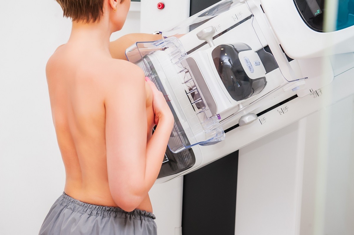
Mammography with tomosynthesis: what it is and what advantages it offers
Mammography with tomosynthesis is a key test for breast cancer prevention, thanks to a more precise diagnosis than ‘classic’ mammography
The resumption of secondary prevention campaigns, which have now returned to pre-pandemic levels, has once again given the female population breathing space, guaranteeing early and early-stage diagnosis of breast cancer, thanks in part to mammography with tomosynthesis.
Mammography with tomosynthesis: what it is, advantages and results
Traditional mammography equipment is gradually being replaced by the mammograph with tomosynthesis.
Tomosynthesis, added in the most modern machines, guarantees a stratigraphic analysis, i.e. a 3D scan of the breast: a millimetric scan of the organ that allows the combination of images captured during the test and ensures a more complete analysis picture.
A new diagnostic means capable of detecting tumour lesions even in its smallest and most circumscribed forms.
The main aim, which is to be achieved through the introduction of these increasingly advanced machines, is the early diagnosis of tumours.
Tomosynthesis makes it possible to reduce false diagnoses and gives us more precise and accurate reports.
In addition, the use of the Mammograph with Tomosynthesis provides further advantages:
- sharper images compared to classic reports
- elimination of artefacts, with more accurate predictive ability;
- less exposure to radiation.
The mammograph with tomosynthesis is particularly effective in young women (40 years old)
Its ability to investigate in depth in the presence of dense mammary glands, (typical of young women,) makes it possible to detect lesions that might once have remained uninterpreted.
The importance of prevention: the return to pre-Covid numbers
With the emergency phase of the Covid pandemic over, we are now witnessing what we can call the side effects of this period.
If before 2019 we were seeing large numbers of women participating in the screening campaigns that the National Health System makes available to them, we now find ourselves with a gap of almost 2 years of mapping.
This gap translates into an increased incidence of oncological diseases and a relative diagnostic delay.
Cancer prevention is therefore today one of the main weapons in the fight against cancer, capable of limiting cancer diseases as far as possible.
The higher the levels of screening coverage, the greater the ability of doctors to tackle the disease in its early stages.
Mammography: the main tool for prevention
The main test for the diagnosis of breast cancer is the mammogram.
This type of diagnostic investigation is fundamental for detecting lesions in the organ and is capable of detecting even the smallest formations.
In fact, Mammography, which is nothing more than an X-ray of the breast, has a high diagnostic power and allows early detection of the disease, helping to intervene early and as effectively as possible on it.
Given its remarkable predictive capacity, screening programmes elect this procedure as their primary test.
Mammography is in fact able to detect possible forms of cancer from their earliest manifestation.
Read Also
Emergency Live Even More…Live: Download The New Free App Of Your Newspaper For IOS And Android
MRI, Magnetic Resonance Imaging Of The Heart: What Is It And Why Is It Important?
Mammary MRI: What It Is And When It Is Done
Mammography: How To Do It And When To Do It
Pap Test: What Is It And When To Do It?
Breast Cancer: For Every Woman And Every Age, The Right Prevention
Transvaginal Ultrasound: How It Works And Why It Is Important
Pap Test, Or Pap Smear: What It Is And When To Do It
Mammography: A “Life-Saving” Examination: What Is It?
Breast Cancer: Oncoplasty And New Surgical Techniques
Gynaecological Cancers: What To Know To Prevent Them
Ovarian Cancer: Symptoms, Causes And Treatment
What Is Digital Mammography And What Advantages It Has
What Are The Risk Factors For Breast Cancer?
Breast Cancer Women ‘Not Offered Fertility Advice’
Ethiopia, The Minister Of Health Lia Taddesse: Six Centers Against Breast Cancer
Breast Self-Exam: How, When And Why
Fusion Prostate Biopsy: How The Examination Is Performed
CT (Computed Axial Tomography): What It Is Used For
What Is An ECG And When To Do An Electrocardiogram
MRI, Magnetic Resonance Imaging Of The Heart: What Is It And Why Is It Important?
Mammary MRI: What It Is And When It Is Done
What Is Needle Aspiration (Or Needle Biopsy Or Biopsy)?
Positron Emission Tomography (PET): What It Is, How It Works And What It Is Used For
CT, MRI And PET Scans: What Are They For?
MRI, Magnetic Resonance Imaging Of The Heart: What Is It And Why Is It Important?
Urethrocistoscopy: What It Is And How Transurethral Cystoscopy Is Performed
What Is Echocolordoppler Of The Supra-Aortic Trunks (Carotids)?
Surgery: Neuronavigation And Monitoring Of Brain Function
Robotic Surgery: Benefits And Risks
Refractive Surgery: What Is It For, How Is It Performed And What To Do?
Single Photon Emission Computed Tomography (SPECT): What It Is And When To Perform It
What Is An ECG And When To Do An Electrocardiogram


