
Melanoma: causes, symptoms, diagnosis and treatment
The incidence of melanoma – until a few years ago considered a rather rare disease – has increased by 4% in the last twenty years, reaching 14.3 cases per 100,000 men and 13.6 cases per 100,000 women
When detected in time and treated appropriately, melanoma is a disease from which it is perfectly possible to recover.
If diagnosed late, however, it can rapidly evolve, invading adjacent tissues and even giving distant metastases, thus becoming fatal.
Melanoma: what is it?
Melanoma is a malignant tumour arising from melanocytes contained mostly in the skin.
Melanoma – an uncommon but often very aggressive tumour – is visible to the naked eye and originates precisely from melanocytes, the cells that produce melanin, the pigment responsible for the colouring of the skin.
Melanoma: causes and symptoms
As already mentioned, melanoma is generated due to the degeneration of melanocytes, the cells responsible for the production of melanin to protect the skin of the entire body from the aggression of ultraviolet rays.
Melanoma can originate either ex-novo or from the degeneration of a pre-existing mole, whose melanocytes undergo degeneration that transforms them into cancerous cells.
Generally, the rate of occurrence of melanoma is increased by incorrect and excessive exposure to sunlight or UV lamps in the absence of adequate sun protection.
For this reason, the areas of the body most likely to be affected by melanoma are those most frequently exposed to the sun: arms, legs, hands and face.
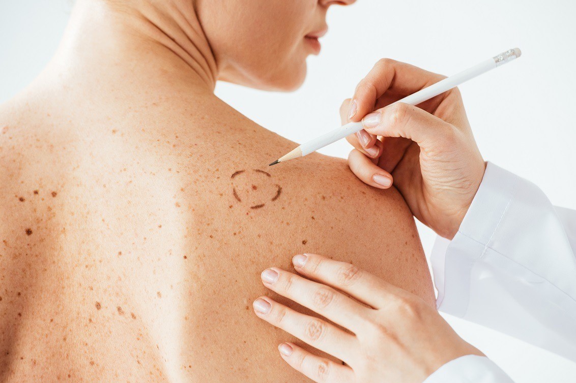
Paying careful attention to the formation of new and existing moles is the first and most important step in diagnosing melanoma early.
The signs to which the most attention should be paid are the following:
- Asymmetry: moles are usually symmetrical, whereas melanoma occurs asymmetrically.
- Irregular edges: Moles have regular, well-defined edges, whereas melanoma has jagged edges.
- Uneven colour: Moles are characterised by a more or less intense but always homogeneous colour, whereas melanoma presents more colours or more gradations of colour.
- Diameter: A suspected skin lesion that is more than 6 millimetres in diameter should be checked by a specialist.
- Evolution: If a mole begins to grow rapidly, change shape or colour this could be a melanoma.
Melanoma: diagnosis
Every individual should spontaneously undergo an annual mole check, during which the dermatologist specialist carries out a careful clinical test.
This allows any moles or suspicious skin spots to be detected, so that the patient can be directed towards specific and targeted tests, such as dermoscopy.
Dermoscopy is a simple and painless procedure that consists of observing moles and skin spots using a specific instrument called an epiluminescence microscope or dermatoscope, hence the name of the test.
This instrument makes it possible to observe the inner appearance of moles, picking out details that would be impossible to detect with the naked eye, thus discovering whether or not the lesion is malignant, so that appropriate action can be taken.
Often, however, observation alone is not enough, so it is necessary to proceed with a histological test by means of a skin biopsy, performed in an outpatient setting.
This is performed by removal – complete or partial – of the lesion, so that a more accurate diagnosis can be made than that allowed by observation alone.
Melanoma staging
The function of the histological test is not only to identify whether or not the portion of tissue removed is a melanoma but – if it is – to determine the staging by localisation of the neoplasm.
The anatomic pathologist, in examining the finding, will in fact be able to determine whether the margins of the resection are healthy or not.
In the former case, it will mean that the melanoma was in locu and that, as it has not yet infiltrated the surrounding tissues, it can be considered completely severed.
Otherwise, by means of well-defined parameters, the anatomic pathologist will be able to predict the depth of invasion of the surrounding tissue by the melanoma, the lymphovascular invasion and the number of multiplying cells, in order to clearly determine the aggressiveness of the melanoma.
If the melanoma is assessed to be aggressive and rapidly growing, the anatomic pathologist will request a biopsy of the sentinel lymph nodes, i.e. those closest to the excised lesion.
If the examination of the sentinel lymph nodes also indicates the presence of cancerous cells, other tests such as ultrasound, X-ray, CT scan and PET scan will be necessary to assess any distant metastases.
Treatment and therapies for melanoma
After assessing and making an accurate diagnosis of the type of tumour we are dealing with, the stage of the disease and the area where it has formed, we proceed with the choice of the most appropriate therapy, which will also depend on other factors such as the patient’s age and state of health.
Surgical therapy
Generally, surgical removal is the recommended treatment for most skin tumours.
With dermatological surgery, the cancerous lesion or suspected cancerous lesion can be completely removed.
Should the removed lesion be large, plastic surgery would intervene to reconstruct the area, especially if it is on the patient’s face, in order to avoid aesthetically compromising it.
When surgery is performed to treat a melanoma, the lymph node responsible for draining the area of skin affected by the removed melanoma – the sentinel lymph node – is usually also removed in order to rule out possible residual cancer cells and prevent distant metastases.
Chemotherapy
Chemotherapy is a treatment consisting of the administration of drugs, either orally or intravenously, that can destroy the cancer cells present.
Skin tumours can also be treated by administering chemotherapy drugs in high concentrations to a limited area, thus preventing the drug from being distributed throughout the body.
Immunotherapy
Immunotherapy is a treatment that makes use of monoclonal antibodies that act by reactivating a part of the immune system that is supposed to specifically and selectively target and destroy tumour cells.
Targeted therapy
Targeted therapies’ refers to particular types of therapies that use drugs to target particular molecular targets of cancer cells, which are considered essential for the growth of the cancer cells themselves.
The characteristic feature of this therapy, as opposed to chemotherapy and radiotherapy, is that it must be tailored to the characteristics of the cancer cell itself.
Melanoma: how can it be prevented?

As mentioned above, the main risk factor for the formation of a melanoma is reckless and irresponsible exposure to ultraviolet rays, both those from natural sources (sunlight) but also and above all those from artificial light (showers or tanning beds).
Ultraviolet rays in fact penetrate the skin and irreparably damage the DNA structure of cells, which may develop mutations that could trigger the development of tumour processes.
To avoid this, prevention is the best cure.
Therefore, avoiding exposure to natural or artificial ultraviolet rays as much as possible, and doing so with due precaution by using high-protection sun creams, is a valid prevention strategy against melanomas or skin cancers in general.
Undergoing an annual mole check with mapping is also recommended for a possible early diagnosis.
In addition, a varied diet rich in vitamins A, C and E – powerful antioxidants – can help the skin protect itself from the harmful action of ultraviolet rays.
Read Also
Emergency Live Even More…Live: Download The New Free App Of Your Newspaper For IOS And Android
Cherry Angiomas: What They Are And How To Remove Them In Minutes
Cavernous Angiomas: What They Are, How To Treat Them
Lymphoma: 10 Alarm Bells Not To Be Underestimated
Non-Hodgkin’s Lymphoma: Symptoms, Diagnosis And Treatment Of A Heterogeneous Group Of Tumours
CAR-T: An Innovative Therapy For Lymphomas
Acute Lymphoblastic Leukaemia: Long-Term Outcomes Described For Childhood ALL Survivors
Lymphangiomas And Lymphatic Malformations: What They Are, How To Treat Them
Melanoma: What Is It And How Can It Be Diagnosed?
Melanoma: Prevention And Dermatological Examinations Are Essential Against Skin Cancer
Nail Melanoma: Prevention And Early Diagnosis
Dermatological Examination For Checking Moles: When To Do It
What Is A Tumour And How It Forms
Rare Diseases: New Hope For Erdheim-Chester Disease
How To Recognise And Treat Melanoma
Moles: Knowing Them To Recognise Melanoma
Skin Melanoma: Types, Symptoms, Diagnosis And The Latest Treatments
Nevi: What They Are And How To Recognise Melanocytic Moles
Bluish Color Of Baby’s Skin: Could Be Tricuspid Atresia
Skin Diseases: Xeroderma Pigmentosum
Basal Cell Carcinoma, How Can It Be Recognised?
Autoimmune Diseases: Care And Treatment Of Vitiligo
Epidermolysis Bullosa And Skin Cancers: Diagnosis And Treatment
SkinNeutrAll®: Checkmate For Skin-Damaging And Flammable Substances
Healing Wounds And Perfusion Oximeter, New Skin-Like Sensor Can Map Blood-Oxygen Levels
Psoriasis, An Ageless Skin Disease
Psoriasis: It Gets Worse In Winter, But It’s Not Just The Cold That’s To Blame
Childhood Psoriasis: What It Is, What The Symptoms Are And How To Treat It
Topical Treatments For Psoriasis: Recommended Over-The-Counter And Prescription Options
What Are The Different Types Of Psoriasis?
Phototherapy For The Treatment Of Psoriasis: What It Is And When It Is Needed
Skin Diseases: How To Treat Psoriasis?
Skin Cancers: Prevention And Care