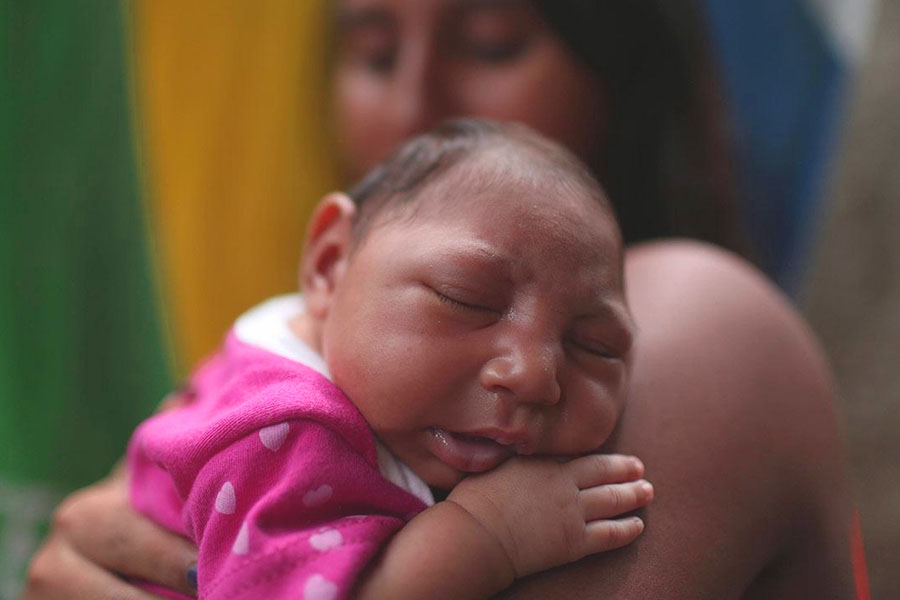
Microcephaly, when the baby's head is much smaller than expected
Microcephaly is a condition characterized by a child’s head circumference being smaller than that of his or her peers and can be generated by both genetic and environmental causes
Microcephaly is caused by a very large and diverse group of diseases
A distinction must be made between genetic causes (e.g., chromosomal abnormalities or single gene abnormalities) and environmental causes.
Microcephaly can be classified into two forms: primary microcephaly (present from birth) and secondary microcephaly (which develops after birth), and may or may not be associated with other symptoms that are part of a more or less characteristic whole (a syndrome).
Microcephaly can be found in more than 450 genetic conditions
The risk of this condition recurring in subsequent parental pregnancies depends on the genetic cause of microcephaly.
In most cases, these are genetic conditions caused by chromosomal aberrations (abnormalities in the number or structure of chromosomes), or by alterations in a single gene, usually inherited in an autosomal recessive manner: both copies of the gene responsible for the disease (the so-called “disease gene”), either the one of maternal or paternal origin, are altered (mutated) in children with microcephaly.
The parents carry an altered copy of the gene (the other copy is normal) and are not affected by microcephaly but are at a 25% chance with each pregnancy of having a child with microcephaly.
Below are the most important forms of microcephaly inherited in an autosomal recessive mode:
Autosomal recessive primary microcephaly
It is characterized by severe microcephaly in the absence of other malformations or neurological defects. The associated cognitive deficit varies from mild to moderate.
MRI scans of patients show a thin but structurally normal cerebral cortex with simplified brain circumvolutions.
The genes most frequently involved in these forms are: ASPM (responsible alone for about 40% of this subtype of microcephaly), MCPH1, PHC1, CENPE, MFSD2A, ANKLE2, CIT, WDFY3, CDKRAP2, CENPJ, STIL, WDR62, CEP63, CEP135, CEP152, CASC5, KNL1, ZNF335, CDK6.
Primordial dwarfism with microcephaly (Seckel syndrome, MOPDI and MOPDII)
This is a group of diseases characterized not only by marked microcephaly but also by poor intrauterine growth and short stature after birth.
At least 9 disease genes are known for Seckel syndrome (CEP63, ATR, NSMCE2, DNA2, CENPJ, NIN, CEP152, RBBP8, TRAIP).
The form called MOPD1 is caused by mutations in the RNU4ATAC gene and the MOPDII form by mutations in the Pericentrin (PCNT) gene.
Smith-Lemli-Opitz syndrome
In some cases microcephaly is inherited in an autosomal dominant mode: it occurs when only one copy of the two disease genes is altered (mutated).
A normal copy of the gene is present, but it is not sufficient to reconstruct the genetic message that has been altered by the mutated gene.
Consequently, a parent who carries a mutated copy of one of the two copies of the gene that can cause microcephaly (and who consequently has microcephaly himself) has a 50% risk, at each conception, of having a child with microcephaly.
In many cases, however, the parents do not have microcephaly and so these are de novo mutations which means that the mutation was not inherited but occurred during the formation of the egg cell, sperm cell, or in the very early stages of embryonic development.
The mutation will then affect only that child and no other family member will be affected.
Microcephaly with congenital lymphedema and chorioretinopathy is inherited as an autosomal dominant condition: the reduction in head circumference is usually mild and is caused by mutation of the KIF11 gene.
Manifestations of the disease variably involve the central nervous system and the eye
The face also has a characteristic appearance with slanted eye slits and protruding chin and ears.
Lymphedema is usually confined to the dorsum of the feet.
Other genetic syndromes associated with microcephaly are caused by more complex genetic mechanisms:
- Angelman syndrome;
- Rubinstein-Taybi syndrome.
The main genetic forms caused by chromosomal aberrations are:
Wolf-Hirschhorn syndrome, caused by a deletion of the short arm of chromosome number 4 (4p16.3).
The deletion leads to loss of a section of the chromosome, thus the genes contained therein.
In 58% of cases, it is such a large deletion that it is detectable under the microscope by karyotype analysis; in the remaining cases, more sophisticated genetic tests (FISH, array-CGH) are needed to prove the deletion.
The clinical picture is characterized by microcephaly, poor growth, intellectual disability, and small deformities,especially in the face, which are quite recognizable to an experienced eye (medical geneticist).
There may be coloboma of the iris (lack of part of the iris presenting in the form of an iris fissure).
Seizures and congenital suffering of the heart muscle are common findings.
During childhood there may be recurrent infections.
Mowat Wilson syndrome
In many cases the cause of microcephaly is not genetic but environmental.
During pregnancy, some environmental causes can cause microcephaly:
- Chemicals such as methylmercury content mainly in fish;
- Narcotic substances and alcohol;
- Infections such as toxoplasmosis, rubella, cytomegalovirus, herpes, chickenpox, HIV, Zika virus;
- Damage done to the brain before birth and during delivery such as hypoxia, ischemia, and trauma;
- Severe malnutrition.
During the examination, the pediatrician, and later the geneticist/neurologist will perform:
- The measurement of the patient’s head circumference, height, and weight, comparing the measurements with special tables showing normal limits for children of the same age and sex; thus it will be possible to assess the extent of microcephaly and discriminate between isolated microcephaly and poor overall growth;
- The measurement of the parents’ head circumference, which is essential to determine whether it is an inherited familial feature;
- Careful review of medical history during pregnancy to rule out environmental causes of microcephaly such as infections, drug use, radiation exposure, travel to countries with endemic infections (e.g., Zika Virus), perinatal asphyxia, and alcohol or drug abuse;
- Assessment of growth at birth, any history of parental consanguinity, progressive or static progression of the condition, psychomotor developmental milestones, any presence of epilepsy, presence or absence of dysmorphic notes, evaluation of any associated malformative abnormalities, and a detailed neurological examination.
Microcephaly is frequently associated with delayed psychomotor development, language, and intellectual disability of varying degrees, but cases in which IQ is within the normal range have also been described.
CHILD HEALTH: LEARN MORE ABOUT MEDICHILD BY VISITING THE BOOTH IN EMERGENCY EXPO
Other symptoms that can occur with microcephaly are:
- Epilepsy;
- Poor growth in stature and weight;
- Anatomical changes in facial shape (facial dysmorphisms);
- Neurobehavioral disorders (hyperactivity, attention deficit, etc.).
The pediatrician, based on evaluation of the family history, analysis of the clinical picture, and examination attempts to identify whether the condition has a genetic basis, in the context of a specific syndrome.
He or she can then propose genetic analysis aimed at identifying, when possible, the cause of microcephaly and calculate accordingly the risk that one of the parents or the couple will again give birth to a child with microcephaly in subsequent pregnancies.
An evaluation by the maxillofacial surgeon to rule out the possibility of craniostenosis is also important.
Neurologic and neuropsychiatric checks are useful because of the possible association between microcephaly and neurologic symptoms such as psychomotor retardation, intellectual disability, and epilepsy.
It is therefore important to perform an electroencephalographic examination.
It may be useful to perform a brain ultrasound and/or MRI to reveal any associated abnormalities of the brain and to assess the status of the cranial sutures.
A fundus examination of the eye may be useful to rule out forms associated with chorioretinopathy.
Ultrasound checks that are routinely performed during pregnancy in some cases may already show a suspicion of microencephaly
However, the prenatal finding must be confirmed through an evaluation during developmental age of skull growth, relating that measurement to those of stature and weight growth.
If a craniostenosis is causing the microcephaly, treatment is surgical.
In other cases, there is no specific therapy.
Multidisciplinary care of the patient according to his or her clinical needs is certainly very important.
In the case of psychomotor retardation and intellectual disability, rehabilitation therapy including psychomotricity, speech therapy, and occupational therapy can be used.
Such therapies have been shown to be effective for the acquisition of some useful skills in daily life and for an improvement in personal autonomy.
If there are seizure-like phenomena, evaluation by the neurologist to set up antiepileptic therapy is indicated.
If other symptoms or organic malformations are present along with microcephaly, the patient should be evaluated by the referring specialists for treatment of the various symptoms.
The prognosis of microcephaly is variable depending on the cause.
The clinical manifestations associated with microcephaly are highly variable.
They range from very mild to severe forms, but generally the prognosis improves over time.
If treatment is adequate, the prognosis of the mild forms of the disease is favorable, and life expectancy is approximately superimposed on that of the general population.
Read Also
Emergency Live Even More…Live: Download The New Free App Of Your Newspaper For IOS And Android
What Is Traumatic Brain Injury (TBI)?
Pediatric Brain Tumors: Types, Causes, Diagnosis And Treatment
Raising The Bar For Pediatric Trauma Care: Analysis And Solutions In The US
Why Are There Leukocytes In My Urine?
Paediatrics / Recurrent Fever: Let’s Talk About Autoinflammatory Diseases
Bone Cysts In Children, The First Sign May Be A ‘Pathological’ Fracture
Foot Deformities: Metatarsus Adductus Or Metatarsus Varus


