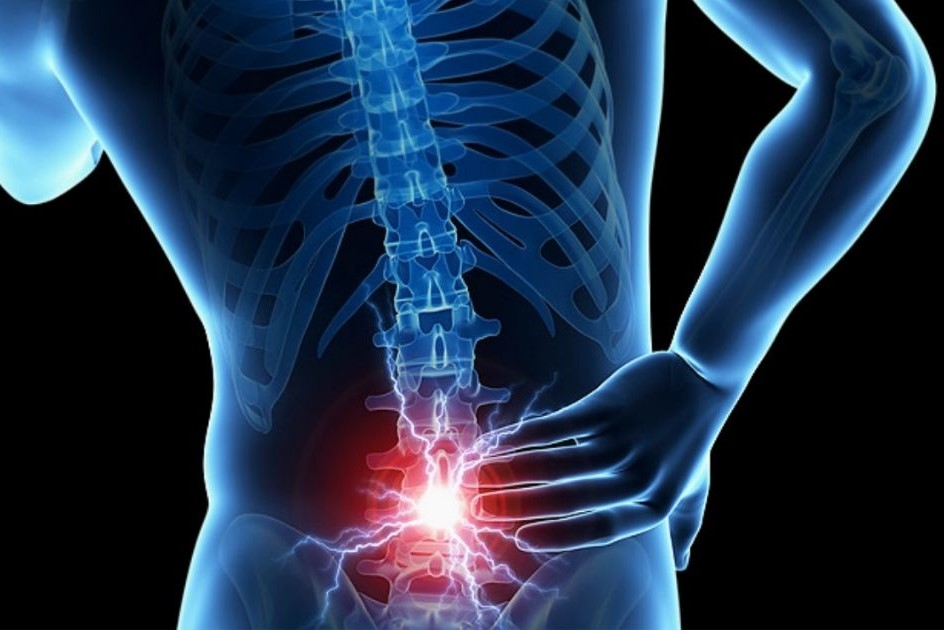
Nerve root disorders: radiculopathies
Radicular pathologies, or radiculopathies, result in predictable segmental root deficits (e.g. pain or paresthesias with a dermatomeric distribution, weakness of muscles innervated by the root)
Diagnosis can be based on a neuroimaging study, electrophysiological tests and general tests to detect possible underlying diseases.
Treatment depends on the cause but includes symptomatic drugs such as NSAIDs, other analgesics and corticosteroids.
Root disorders (radiculopathies) are caused by an acute or chronic overload of pressure on a nerve root within the adjacent area of the spine
Aetiology of nerve root diseases
The most frequent cause of radiculopathies is
- A herniated intervertebral disc
Bone changes due to rheumatoid arthritis or arthrosis, especially when localised in the cervical or lumbar region, can also lead to compression of isolated nerve roots.
Less frequently, meningeal carcinomatosis causes multiple multi-segmental root dysfunction.
Rarely, spinal masses (e.g. epidural abscesses and tumours, spinal meningiomas, neurofibromas) may manifest with radicular symptoms instead of the usual symptoms of spinal cord dysfunction.
Diabetes may cause painful radiculopathy in the chest or extremities due to nerve root ischaemia.
Infectious diseases, such as those caused by mycobacteria (e.g. tuberculosis), fungi (e.g. histoplasmosis) or spirochete diseases (e.g. Lyme disease, syphilis), sometimes affect the nerve roots.
Shingles infection usually causes a painful radiculopathy with loss of sensation with dermatomeric distribution and a characteristic rash, but may cause a motor radiculopathy with segmental weakness and loss of reflexes.
Cytomegalovirus-induced polyradiculitis is a complication of AIDS.
Symptomatology of nerve root disorders (radiculopathies)
Radiculopathies tend to cause a characteristic painful radicular syndrome and segmental neurological deficits depending on the medullary level corresponding to the affected root.
The muscles innervated by the affected motor root become weak and atrophic; they may also become flaccid with fasciculations.
Involvement of the sensory root causes sensory impairments with a dermatomeric distribution.
The corresponding segmental osteotendinous reflexes may be reduced or absent.
Electric shock-like pain may radiate along the distribution of the affected nerve root.
Pain may be exacerbated by movements that transmit pressure to the nerve root through the subarachnoid space (e.g., moving the spine, coughing, sneezing, doing the Valsalva manoeuvre).
Lesions of the cauda equina, affecting multiple sacral and lumbar roots, produce radicular symptoms in both legs and may induce sphincter and sexual function alterations.
Evidence suggestive of spinal cord compression is as follows:
- Presence of a sensory level (an abrupt change in sensation below a dermatomere, i.e. below a horizontal line passing through the spinal cord, at a specific level)
- Paraparesis or flaccid tetraparesis
- Alterations in reflexes below the site of compression
- Hyporeflexia at onset, later followed by hyperreflexia
- Sphincter dysfunction
Diagnosis of nerve root pathologies
- Neuroimaging
- Sometimes electrophysiological examinations
The presence of radicular symptoms requires an MRI or CT scan of the affected area.
Myelography is only necessary if MRI is contraindicated (e.g. due to a pacemaker or the presence of other metals) and if CT is inconclusive.
The area studied depends on the symptomatology; if the level is unclear, electrophysiological tests should be carried out to localise the affected roots, although they do not distinguish between the various causes.
If neuroimaging does not detect an anatomical abnormality, cerebrospinal fluid analysis is performed to rule out an infectious or inflammatory cause and fasting plasma glucose (FPG) is measured to check for diabetes.
Treatment of nerve root disorders (radiculopathies)
- Treatment of the cause and pain
- Surgery (as a last resort)
The specific causes of nerve root diseases are treated.
Acute pain requires and appropriate analgesics (e.g. paracetamol, non-steroidal anti-inflammatory drugs, sometimes opioids).
NSAIDs are particularly useful for diseases involving inflammation.
Myorelaxants, hypnotics and topical treatments rarely provide additional benefit.
If symptoms are not significantly relieved with non-opioid analgesics, corticosteroids may be administered systemically or as an epidural injection; however, analgesia tends to be modest and temporary.
Methylprednisolone can be used, with subsequent gradual scaling up over 6 days, starting at 24 mg orally once/day and decreasing by 4 mg/day.
The management of chronic pain can be difficult; acetaminophen (paracetamol) and NSAIDs are often only partially effective, and long-term use of NSAIDs carries considerable risks.
Opioids have a high risk of addiction.
Tricyclic antidepressants and anticonvulsants can be effective, as can physiotherapy and a medical examination with a psychiatrist.
For a few patients, alternative medical treatments (e.g. transdermal nerve stimulation, spinal manipulation, acupuncture, herbal medicines) may be tried if all others are ineffective.
If pain is intractable or if progressive sphincter weakness or dysfunction suggests spinal compression, surgical decompression may be required.
Read Also:
Emergency Live Even More…Live: Download The New Free App Of Your Newspaper For IOS And Android
O.Therapy: What It Is, How It Works And For Which Diseases It Is Indicated
Oxygen-Ozone Therapy In The Treatment Of Fibromyalgia
When The Patient Complains Of Pain In The Right Or Left Hip: Here Are The Related Pathologies
Why Do Muscle Fasciculations Occur?


