Neurogenic shock: what it is, how to diagnose it and how to treat the patient
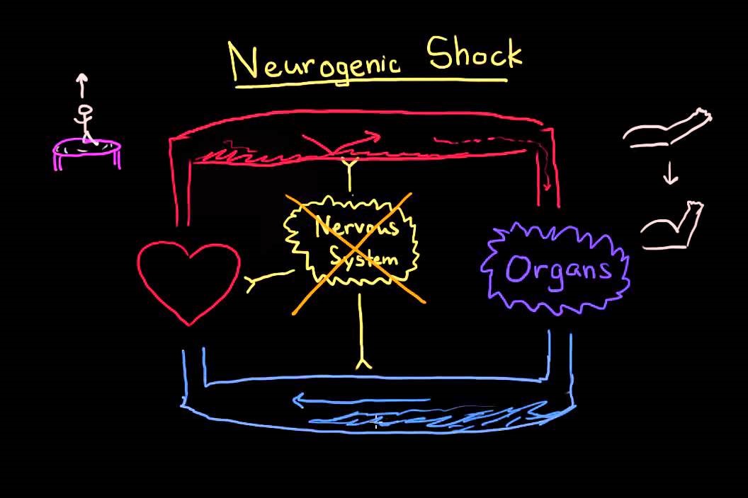
In neurogenic shock, vasodilation occurs as a result of a loss of balance between parasympathetic and sympathetic stimulation
What is Neurogenic Shock?
Neurogenic shock is a distributive type of shock.
In neurogenic shock, vasodilation occurs as a result of a loss of balance between parasympathetic and sympathetic stimulation.
It is a type of shock (a life-threatening medical condition in which there is insufficient blood flow throughout the body) that is caused by the sudden loss of signals from the sympathetic nervous system that maintain the normal muscle tone in blood vessel walls.
The patient experiences the following that results in neurogenic shock:
- Stimulation. Sympathetic stimulation causes vascular smooth muscle to constrict, and parasympathetic stimulation causes vascular smooth muscle to relax or dilate.
- Vasodilation. The patient experiences a predominant parasympathetic stimulation that causes vasodilation lasting for an extended period of time, leading to a relative hypovolemic state.
- Hypotension. Blood volume is adequate, because the vasculature is dilated; the blood volume is displaced, producing a hypotensive (low BP) state.
- Cardiovascular changes. The overriding parasympathetic stimulation that occurs with neurogenic shock causes a drastic decrease in the patient’s systemic vascular resistance and bradycardia.
- Insufficient perfusion. Inadequate BP results in the insufficient perfusion of tissues and cells that is common to all shock states.
THE RADIO OF THE WORLD’S RESCUERS? VISIT THE RADIO EMS BOOTH AT EMERGENCY EXPO
Neurogenic shock could be caused by the following:
- Spinal cord injury. Spinal cord injury (SCI) is recognised to cause hypotension and bradycardia (neurogenic shock).
- Spinal anesthesia. Spinal anesthesia—injection of an anesthetic into the space surrounding the spinal cord—or severance of the spinal cord results in a fall in blood pressure because of dilation of the blood vessels in the lower portion of the body and a resultant diminution of venous return to the heart.
- Depressant action of medications. Depressant action of medications and lack of glucose could also cause neurogenic shock.
The clinical manifestations of neurogenic shock are signs of parasympathetic stimulation
- Dry, warm skin. Instead of cool, moist skin, the patient experiences dry, warm skin due to vasodilation and inability to vasoconstrict.
- Hypotension. Hypotension occurs due to sudden, massive dilation.
- Bradycardia. Instead of getting tachycardic, the patient experience bradycardia.
- Diaphragmatic breathing. If the injury is below the 5th cervical vertebra, the patient will exhibit diaphragmatic breathing due to loss of nervous control of the intercostal muscles (which are required for thoracic breathing).
- Respiratory arrest. If the injury is above the 3rd cervical vertebra, the patient will go into respiratory arrest immediately following the injury, due to loss of nervous control of the diaphragm.
TRAINING: VISIT THE BOOTH OF DMC DINAS MEDICAL CONSULTANTS IN EMERGENCY EXPO
Assessment and Diagnostic Findings
Diagnosis of neurogenic shock is possible through the following tests:
- Computerized tomography (CT) scan. A CT scan may provide a better look at abnormalities seen on an X-ray.
- Xrays. Medical personnel typically order these tests on people who are suspected of having a spinal cord injury after trauma.
- Magnetic resonance imaging (MRI). MRI uses a strong magnetic field and radio waves to produce computer-generated images.
Medical Management
Treatment of neurogenic shock involves:
- Restoring sympathetic tone. It would be either through the stabilization of a spinal cord injury or, in the instance of spinal anesthesia, by positioning the patient appropriately.
- Immobilization. If the patient has a suspected case of spinal cord injury, a traction may be needed to stabilize the spine to bring it to proper alignment.
- IV fluids. Administration of IV fluids is done to stabilize the patient’s blood pressure.
Pharmacologic Therapy
Drugs administered to a patient undergoing neurogenic shock are:
- Inotropic agents. Inotropic agents such as dopamine may be infused for fluid resuscitation.
- Atropine. Atropine is given intravenously to manage severe bradycardia.
- Steroids. Patient with obvious neurological deficit can be given I.V. steroids, such as methylprednisolone in high dose, within 8 hours of commencement of neurogenic shock.
- Heparin. Administration of heparin or low molecular-weight heparin as prescribed may prevent thrombus formation.
Nursing management of a patient with neurogenic shock includes:
Nursing Assessment
Assessment of a patient with neurogenic shock should involve:
- ABC assessment. The prehospital provider should follow the basic airway, breathing, circulation approach to the trauma patient while protecting the spine from any extra movement.
- Neurologic assessment. Neurologic deficits and a general level at which abnormalities began should be identified.
Nursing Diagnosis
Based on the assessment data, the nursing diagnoses for a patient with neurogenic shock are:
- Risk for impaired breathing pattern related to impairment of innervation of diaphragm (lesions at or above C-5).
- Risk for trauma related to temporary weakness/instability of spinal column.
- Impaired physical mobility related to neuromuscular impairment.
- Disturbed sensory perception related to destruction of sensory tracts with altered sensory reception, transmission, and integration.
- Acute pain related to pooling of the blood secondary to thrombus formation.
Nursing Care Planning & Goals
The major goals for the patient include:
- Maintain adequate ventilation as evidenced by absence of respiratory distress and ABGs within acceptable limits
- Demonstrate appropriate behaviors to support the respiratory effort.
- Maintain proper alignment of spine without further spinal cord damage.
- Maintain position of function as evidenced by absence of contractures, foot drop.
- Increase strength of unaffected/compensatory body parts.
- Demonstrate techniques/behaviors that enable resumption of activity.
- Recognize sensory impairments.
- Identify behaviors to compensate for deficits.
- Verbalize awareness of sensory needs and potential for deprivation/overload.
Nursing Interventions
- Nursing interventions are directed towards supporting cardiovascular and neurologic function until the usually transient episode of neurogenic shock resolves.
- Elevate head of bed. Elevation of the head helps prevent the spread of the anesthetic agent up the spinal cord when a patient receives spinal or epidural anesthesia.
- Lower extremity interventions. Applying anti-embolism stockings and elevating the foot of the bed may help minimize pooling of the blood in the legs and prevent thrombus formation.
- Exercise. Passive range of motion of the immobile extremities helps promote circulation.
- Airway patency. Maintain patent airway: keep head in neutral position, elevate head of bed slightly if tolerated, use airway adjuncts as indicated.
- Oxygen. Administer oxygen by appropriate method (nasal prongs, mask, intubation, ventilator).
- Activities. Plan activities to provide uninterrupted rest periods and encourage involvement within individual tolerance and ability.
- BP monitoring. Measure and monitor BP before and after activity in acute phases or until stable.
- Reduce anxiety. Assist patient to recognize and compensate for alterations in sensation.
Evaluation
Expected patient outcomes are:
- Maintained adequate ventilation.
- Demonstrated appropriate behaviors to support the respiratory effort.
- Maintained proper alignment of spine without further spinal cord damage.
- Maintained position of function.
- Increased strength of unaffected/compensatory body parts.
- Demonstrated techniques/behaviors that enable resumption of activity.
- Recognized sensory impairments.
- Identified behaviors to compensate for deficits.
- Verbalized awareness of sensory needs and potential for deprivation/overload.
Documentation Guidelines
The focus of documentation are:
- Relevant history of problem.
- Respiratory pattern, breath sounds, use of accessory muscles.
- Laboratory values.
- Past and recent history of injuries, awareness of safety needs.
- Use of safety equipment or procedures.
- Environmental concerns, safety issues.
- Level of function, ability to participate in specific or desired activities.
- Client’s description of response to pain, specifics of pain inventory, expectations of pain management, and acceptable level of pain.
- Prior medication use.
- Plan of care, specific interventions, and who is involved in the planning.
- Teaching plan.
- Response to interventions, teaching, actions performed, and treatment regimen.
- Attainment or progress towards desired outcomes.
- Modifications to the plan of care.
Read Also
Emergency Live Even More…Live: Download The New Free App Of Your Newspaper For IOS And Android
Circulatory Shock (Circulatory Failure): Causes, Symptoms, Diagnosis, Treatment
The Quick And Dirty Guide To Shock: Differences Between Compensated, Decompensated And Irreversible
Cardiogenic Shock: Causes, Symptoms, Risks, Diagnosis, Treatment, Prognosis, Death
Anaphylactic Shock: What It Is And How To Deal With It
Basic Airway Assessment: An Overview
Respiratory Distress Emergencies: Patient Management And Stabilisation
Behavioural And Psychiatric Disorders: How To Intervene In First Aid And Emergencies
Fainting, How To Manage The Emergency Related To Loss Of Consciousness
Altered Level Of Consciousness Emergencies (ALOC): What To Do?
Syncope: Symptoms, Diagnosis And Treatment
How Healthcare Providers Define Whether You’re Really Unconscious
Cardiac Syncope: What It Is, How It Is Diagnosed And Who It Affects
New Epilepsy Warning Device Could Save Thousands Of Lives
Understanding Seizures And Epilepsy
First Aid And Epilepsy: How To Recognise A Seizure And Help A Patient
Neurology, Difference Between Epilepsy And Syncope
First Aid And Emergency Interventions: Syncope
Epilepsy Surgery: Routes To Remove Or Isolate Brain Areas Responsible For Seizures
Trendelenburg (Anti-Shock) Position: What It Is And When It Is Recommended
Head Up Tilt Test, How The Test That Investigates The Causes Of Vagal Syncope Works


