Nuclear Magnetic Resonance (NMR): when to do it?
Nuclear Magnetic Resonance (NMR) is a physical phenomenon characteristic of nuclei exposed to a magnetic field
When applied to the medical field, it is called TRM (Magnetic Resonance Tomography) or more simply MRI.
In this case, it is a diagnostic technique based on the use of a magnetic field and radiofrequency electromagnetic waves.
Nuclear Magnetic Resonance (NMR) provides detailed images of the human body
With this technique, many diseases and changes in the internal organs can be visualised and thus easily diagnosed.
With MRI, soft tissues are clearly visible and discrimination between tissue types is possible, which is sometimes not appreciable with other radiological techniques.
MRI is, from a technological point of view, much more recent than CT and is still in full evolution.
It is a harmless examination that uses neither X-rays nor radioactive sources, although in some cases (such as in pregnant patients) it can be considered potentially harmful and is only used after careful risk/benefit assessment.
What Nuclear Magnetic Resonance Imaging (NMR) consists of
- The patient is made to lie supine on a couch.
- Depending on the type of organ to be studied, so-called ‘surface coils’ (helmet, bands, plates, etc.) shaped to fit the anatomical region concerned may be placed on the outside of the body;
- the application of these ‘coils’ causes no pain or discomfort to the patient.
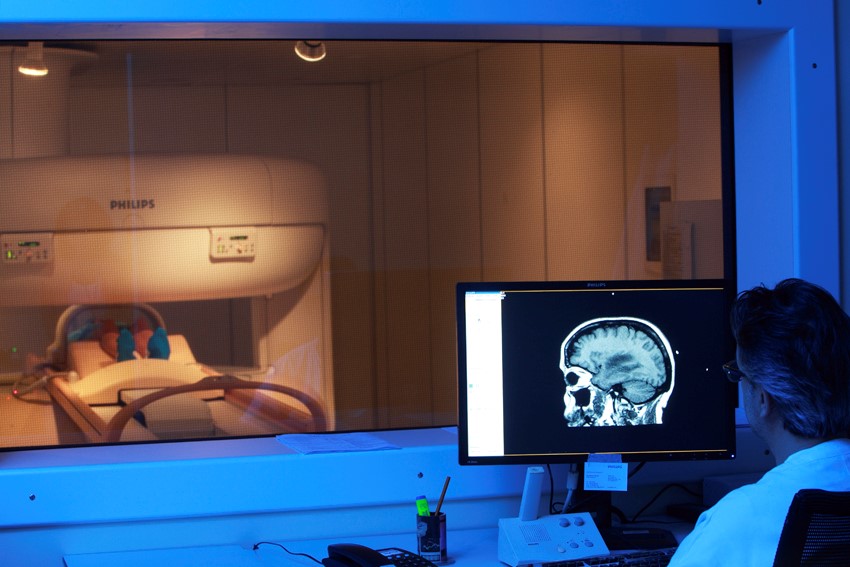
- The patient is introduced inside the MRI machine, a fairly large and comfortable tube, open at both ends and with equipment that allows communication with the personnel in charge of conducting the examination.
- In this machine, he is irradiated by a high-intensity magnetic field.
- The patient must remain motionless for the duration of the examination.
Inside the machine, the forces generated in the magnetic field cause the magnetic moments of the patient’s molecules to align with the direction of the external field, inducing temporary alterations in the nuclei, which, when the radio waves are interrupted, return to normal, resulting in signals.
The signals are then transmitted to a computer and transformed into three-dimensional images.
In these images, tissues are light-coloured if they are rich in water, due to the abundant presence of hydrogen atoms (the basic element of biological tissues), and dark if they are poor in water.
If images are acquired in rapid sequence, they will also allow the visualisation of films, e.g. of cardiac motion or the accumulation of contrast medium in tissues.
Images can also be printed on radiographic-like film.
There are no radiation risks and, therefore, the investigation is safe, painless and essentially free of side effects.
Nuclear Magnetic Resonance has a variable duration, but on average the time spent inside the machine is about 30 minutes
Once the diagnostic examination is over, the patient can go home without any particular problems.
During the MRI, at the radiologist’s discretion and depending on the type of pathology to be studied, an intravenous contrast medium may be administered.
Unlike other diagnostic investigations (e.g. angiography or CT scan), the amount of contrast medium generally required for diagnosis is relatively small (10-20 ml).
The use of contrast medium has no side effects, apart from rare cases of allergic reaction.
Recently, an influence of paramagnetic contrast medium on the emergence of a syndrome called systemic nephrogenic fibrosis in patients with severe, acute or chronic renal failure or with renal dysfunction due to hepato-renal syndrome in the perioperative period
These cases are very rare and there are adequate control and protection protocols.
Before the examination, the patient must remove all metal objects (watch, glasses, hairpins, jewellery, etc.) and clothing that may contain fibres or metal parts (corsets, bodysuit bras, etc.); they must also remove all cosmetic products and dentures. In general, no special preparations or diets should be followed.
When and why Nuclear Magnetic Resonance Imaging is used
Magnetic Resonance Imaging represents the most modern imaging method available today and, therefore, can be used to diagnose a wide variety of pathological conditions involving the body’s organs and tissues.
MRI is useful in the diagnosis of diseases of the brain and spine, abdomen and pelvis (liver and uterus), large vessels (aorta) and the musculoskeletal system (joints, bone, cartilage).
It is particularly useful for the study of soft tissues (muscles, blood vessels, liver, ligaments, nervous system, heart and all internal organs), which are rich in water and thus in hydrogen atoms, and less so for the examination of ‘hard’ anatomical structures, which are deficient in water (bone).
MRI is contraindicated for patients who are pregnant or wearers of pace-makers, metal heart valves, prostheses with electronic circuits and metal preparations placed near vital organs.
The indications for the use of MRI are evolving, not least because of the more recent introduction of new CT techniques that allow findings that were unthinkable just a few years ago.
It is therefore important to be aware that MRI is not always the best examination; there are cases in which MRI and CT have overlapping results, and cases in which CT is preferable (e.g. study of osteodiscal pathology in the elderly).
The radiologist is able to indicate the preferable examination on a case-by-case basis.of liver transplantation.
Read Also
Emergency Live Even More…Live: Download The New Free App Of Your Newspaper For IOS And Android
Medical Thermography: What Is It For?
Positron Emission Tomography (PET): What It Is, How It Works And What It Is Used For
Single Photon Emission Computed Tomography (SPECT): What It Is And When To Perform It
Instrumental Examinations: What Is The Colour Doppler Echocardiogram?
Coronarography, What Is This Examination?
CT, MRI And PET Scans: What Are They For?
MRI, Magnetic Resonance Imaging Of The Heart: What Is It And Why Is It Important?
Urethrocistoscopy: What It Is And How Transurethral Cystoscopy Is Performed
What Is Echocolordoppler Of The Supra-Aortic Trunks (Carotids)?
Surgery: Neuronavigation And Monitoring Of Brain Function
Robotic Surgery: Benefits And Risks
Refractive Surgery: What Is It For, How Is It Performed And What To Do?


