Pathological anatomy and pathophysiology: neurological and pulmonary damage from drowning
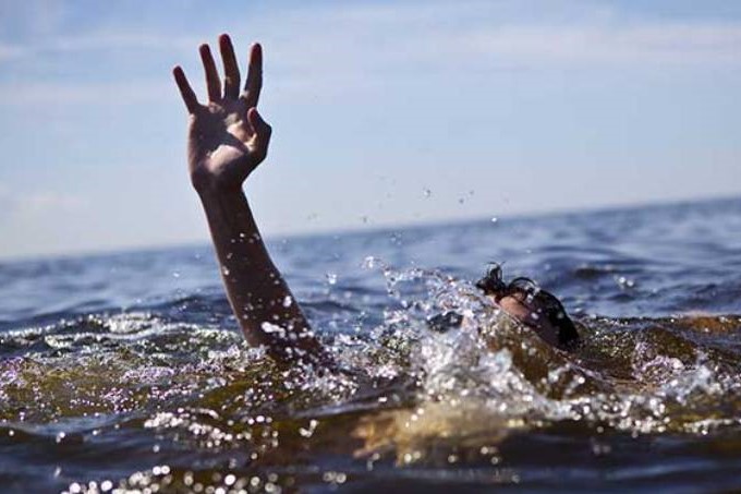
Drowning or ‘drowning syndrome’ in medicine refers to a form of acute asphyxia from an external mechanical cause caused by the occupation of the pulmonary alveolar space by water or other liquid introduced through the upper airways, which are completely submerged in such liquid
If the asphyxia is prolonged for a long time, usually several minutes, ‘death by drowning’ occurs, i.e. death due to suffocation by immersion, generally linked to acute hypoxia and acute failure of the right ventricle of the heart.
In some non-fatal cases, drowning can be successfully treated with specific resuscitation manoeuvres
The concepts of hypoxia, ischaemia and necrosis are important and must be clarified in detail.
Hypoxia is defined as an inadequate supply of oxygen to a specific body district.
Ischaemia occurs when the blood flow to an organ or apparatus is reduced, or when blood oxygen levels are significantly lower than normal: in these cases, if the blood flow is not restored quickly, the tissue may go into necrosis, i.e. die.
In the case of failure to drown, the brain may become hypoxic before cardiac arrest occurs.
Blood flow may continue for some time, under anaerobic conditions, even after complete consumption of available oxygen.
In most cases, loss of consciousness occurs after 2 minutes of anoxia, and brain damage may occur after 4-6 minutes; nerve damage in some cases is irreversible.
There is no real time limit for recovery, as this depends on numerous factors: cases of complete recovery after periods of immersion lasting up to 40 minutes have been described.
These exceptional cases are more frequent when the accident occurs in cold water, and can be explained by the integrity of the diving reflex (apnoea, bradycardia and peripheral vasoconstriction when the face is immersed in cold water).
Probably the rapid onset of hypothermia, by reducing metabolic demands, especially encephalic ones, exerts cerebro-protective effects and thus contributes to a greater possibility of functional recovery even after many minutes.
Under aerobic conditions, energy production in the form of adenosine triphosphate (ATP) occurs through metabolic pathways such as glycolysis, the tricarboxylic acid cycle (TCA) and oxidative phosphorylation.
There are four important metabolic stages:
Phase I: digestion and absorption of fats, carbohydrates and proteins.
Phase II: Reduction of fatty acids, glucose and amino acids to acetyl-coenzyme A (acetyl=coA), which can be used, as required, either to synthesise fats, carbohydrates or amino acids again, either directly or indirectly, or to obtain additional energy through phases III and IV.
Phase III: Tricarboxylic acid cycle, in which most of the organism’s carbon dioxide (CO2) is produced and in which most of the molecular energy carriers (nicotinamide-adenine dinucleotide [NAD], flavin-adenine dinucleatide [FAD]) take up their energy content (in the form of hydrogen atoms). These carriers transport energy to the respiratory chain.
Phase IV: oxidative phosphorylation (production of adenosine triphosphate [ATP] in the presence of oxygen) takes place at the inner mitochondrial membrane, with oxygen being the final acceptor of the electrons now depleted of energy content and hydrogen atoms.
Glycolysis takes place in the cytoplasm, while the TCA cycle and oxidative phosphorylation occur within the mitochondria.
In anaerobiosis, the TCA cycle and oxidative phosphorylation stop, and the main source of energy remains glycolysis.
Glycolysis, under anaerobic conditions, is rapid but requires the maintenance of blood flow, which is necessary to ensure glucose supply.
Anaerobic metabolism of a glucose molecule results in the net production of 2 ATP molecules, compared to the 36 produced in aerobiosis.
ATP provides the energy for many active transport mechanisms (sodium-potassium pumps, calcium pumps, etc.) present on cell membranes and necessary for maintaining homeostasis.
Brain cells have a strictly aerobic metabolism and, under hypoxic conditions, can rapidly be compromised by a reduction in oxygen and energy supply, which results in a slowdown or complete shutdown of active transport mechanisms.
The integrity of cellular structures is jeopardised by the loss of potassium across the plasma membrane and the influx of sodium and calcium into the cells.
Mitochondria and the endoplasmic reticulum (ER) are intracellular organelles that cooperate in the regulation of cytoplasmic calcium levels, absorbing it when in excess.
Under hypoxic conditions, when cellular integrity begins to be compromised, calcium uptake by these organelles is the proximate cause of the uncoupling of oxidative phosphorylation, a phenomenon that greatly reduces energy production and further deteriorates cellular metabolism.
Water follows sodium and calcium into the cells, leading to oedema.
The end product of the glycolytic pathway is pyruvate under aerobic conditions, and lactate (lactic acid) under aerobic conditions.
The accumulation of lactate reduces pH and can impair the functionality of enzyme systems, leading to cell death if oxygenation and perfusion are not restored.
Pathological anatomy and pathophysiology: drowning lung damage
Fluid aspiration (wet drowning) occurs in approximately 85-90% of drowning victims.
Lung injuries occur more frequently in this group than in patients who have not aspirated.
The extent of these injuries depends on the volume and type of fluid aspirated, as well as any substances contained in it.
The difference between drowning in salt or fresh water is important:
- fresh water is hypotonic compared to blood and, if sucked in, is rapidly absorbed into the circulation. It also destroys surfactant, thereby increasing the surface tension at the level of the alveoli, leading to their collapse;
- seawater is hypertonic with respect to the blood (saline solution at around 3%) and, if sucked in, draws fluid from the blood into the alveoli. This results, in succession, in the mechanical removal of surfactant, increased superficial tension and alveolar collapse.
Atelectasis results in a disaccommodation of the ventilation-perfusion ratio (V/Q), an intrapulmonary shunt (Qs/Qt), a reduction in residual functional capacity and a reduction in lung compliance.
These alterations often result in transient hypoxaemia.
Mixed with fluid, mud, sand, bacteria and gastric material may be aspirated, which are responsible for inflammatory processes in the airways, such as alveolitis, bronchitis and pneumonia.
ARDS is a frequent complication of failed drowning cases, and most likely results from microvascular injury associated with the aspiration of foreign materials and/or the inflammatory response triggered by them.
Activated granulocytes release lysosomal enzymes and oxygen free radicals, and may damage the alveoli-capillary membrane, causing protein-rich fluid to flow into the interstitial spaces, from where it is very difficult for it to be removed.
The adhesion of protein material to the alveolar walls can lead to the formation of hyaline membranes, to which corresponds the whitish appearance on chest X-ray, characteristic of ARDS.
ARDS, once realised, resolves very slowly.
Pathology and pathophysiology: haemodynamic and electrolyte effects
Animal studies have shown no difference between hypoxic animals and animals that were given hypotonic, isotonic or hypertonic saline.
Pulmonary vascular resistance, central venous pressure and pulmonary capillary wedge pressure increased in all animals, while cardiac output and effective dynamic pulmonary compliance decreased.
An equally important finding was the absence of significant haemodynamic or cardiovascular differences between the hypoxic control subjects and those aspirating the various solutions.
Functional, haemodynamic and cardiovascular changes appear more easily during hypoxia than during fluid aspiration.
The study of drowning victims, whether in fresh or salt water, did not document serious alterations in haemoglobin or electrolyte concentrations.
Consequently, haemoglobin and haematocrit values do not make it possible to determine whether fresh or salt water was aspirated.
Pathological anatomy and pathophysiology: damage to renal function in victims of drowning failure
Most victims of a near-drowning do not experience renal function impairment, however, it does occur in some cases and should not be underestimated.
Acute tubular necrosis may be due to myoglobinuria, reduced renal blood flow secondary to the hypoxic event, hypotension, lactic acid production, trauma.
Maintaining an adequate cardiac output is usually sufficient to prevent the onset of renal failure.
Read Also
Emergency Live Even More…Live: Download The New Free App Of Your Newspaper For IOS And Android
Drowning: Symptoms, Signs, Initial Assessment, Diagnosis, Severity. Relevance Of The Orlowski Score
Emergency Interventions: The 4 Stages Preceding Death By Drowning
First Aid: Initial And Hospital Treatment Of Drowning Victims
First Aid For Dehydration: Knowing How To Respond To A Situation Not Necessarily Related To The Heat
Children At Risk Of Heat-Related Illnesses In Hot Weather: Here’s What To Do
Dry And Secondary Drowning: Meaning, Symptoms And Prevention
Drowning In Salt Water Or Swimming Pool: Treatment And First Aid
Drowning Resuscitation For Surfers
Risk Of Drowning: 7 Swimming Pool Safety Tips
First Aid In Drowning Children, New Intervention Modality Suggestion
Water Rescue Dogs: How Are They Trained?
Drowning Prevention And Water Rescue: The Rip Current
Water Rescue: Drowning First Aid, Diving Injuries
RLSS UK Deploys Innovative Technologies And The Use Of Drones To Support Water Rescues / VIDEO
Civil Protection: What To Do During A Flood Or If A Inundation Is Imminent
Floods And Inundations, Some Guidance To Citizens On Food And Water
Emergency Backpacks: How To Provide A Proper Maintenance? Video And Tips
Civil Protection Mobile Column In Italy: What It Is And When It Is Activated
Disaster Psychology: Meaning, Areas, Applications, Training
Medicine Of Major Emergencies And Disasters: Strategies, Logistics, Tools, Triage
Floods And Inundations: Boxwall Barriers Change The Scenario Of The Maxi-Emergency
Disaster Emergency Kit: how to realize it
Earthquake Bag : What To Include In Your Grab & Go Emergency Kit
Major Emergencies And Panic Management: What To Do And What NOT To Do During And After An Earthquake
Earthquake And Loss Of Control: Psychologist Explains The Psychological Risks Of An Earthquake
Earthquake and How Jordanian hotels manage safety and security
PTSD: First responders find themselves into Daniel artworks
Emergency preparedness for our pets


