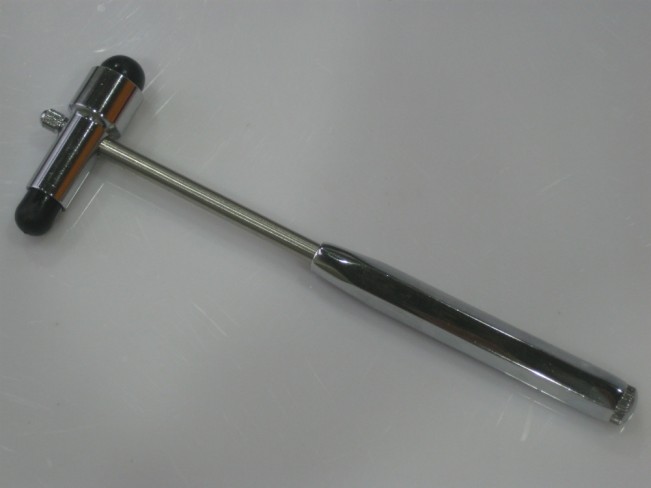
Patient conditions: how to assess reflexes
Assessment of reflexes, whether pupillary or tendon reflexes, is an essential step in determining the condition of the patient you are treating or rescuing
Deep tendon reflexes
Eliciting osteotendinous (muscle stretch) reflexes assesses afferent nerves, synaptic connections within the spinal cord, motor nerves and descending motor pathways.
Lower motor neuron lesions (e.g., those affecting the anterior horn cells, spinal roots, or peripheral nerves) depress reflexes; upper motor neuron lesions (i.e., at any level above the anterior horn cells, except for basal ganglia lesions) increase reflexes.
Reflexes tested include:
- Biceps (innervated by C5 and C6)
- Radiobrachial (from C6)
- Triceps (from C7)
- Distal finger flexors (from C8)
- Quadriceps knee reflex (from L4)
- Achilles tendon reflex (from S1)
- Mandibular reflex (from the V cranial nerve)
It is necessary to note any asymmetry either in the sense of an increase or a reduction.
The Jendrassik manoeuvre can be used to increase hypoactive reflexes: the patient intertwines their hands together and exerts traction (as if to separate them) while a tendon in the lower limb is struck with the hammer.
Alternatively, the patient can push their knees against each other while a tendon in the upper limb is tested.
Pathological reflexes
Pathological reflexes (e.g. Babinski’s, Chaddock’s, Oppenheim’s, snout reflex, sucking and prehension reflex) represent a regression to primitive responses and indicate a loss of cortical inhibition.
Babinski’s, Chaddock’s and Oppenheim’s reflexes all assess plantar response
The normal reflex response is flexion of the big toe.
An abnormal response is slower and consists of extension of the big toe with flaring of the other toes and often flexion of the knee and hip.
This response is of spinal reflex origin and indicates a lack of spinal inhibition due to an upper motor neuron lesion.
For the Babinski reflex, the lateral region of the sole of the foot from the heel to the forefoot is stimulated forcefully using a tongue depressor or the blunt end of a reflex hammer.
The stimulus should be consistent but not injurious; the manoeuvre should not be performed too medially as it may inadvertently induce a primitive prehension reflex.
In sensitive individuals, the reflex response may be masked by rapid withdrawal of the foot, which is not a problem when testing the Chaddock or Oppenheim reflex.
For the Chaddock reflex, the lateral side of the foot, from the lateral malleolus to the little toe, is stimulated with a blunt instrument.
For the Oppenheim reflex, the examiner rubs the anterior tibial side firmly with the knuckles, from just below the kneecap to the foot.
The Oppenheim test can be used with the Babinski test or the Chaddock test to make withdrawal less likely.
The muzzle reflex is present if drumming a tongue depressor between the lips evokes lip protrusion.
The seeking reflex is present if rubbing the upper lip in the lateral portion evokes a movement of the mouth towards the stimulus.
The prehension reflex is present when gentle stimulation of the patient’s palm causes the fingers to flex and grasp the examiner’s finger.
The palmomenton reflex is present if rubbing the palm of the hand evokes contraction of the ipsilateral mental muscle of the lower lip.
Hoffmann’s sign is present if lightly tapping down on the nail of the third or fourth finger evokes an involuntary flexion of the distal phalanx of the thumb and index finger.
Tromner’s sign is similar to Hoffman’s sign, but the finger is struck upwards.
For the glabella sign, one drums on the forehead to induce the blink; normally, each of the first 5 touches induces a single blink, then the reflex is extinguished.
Blinking persists in patients with diffuse brain dysfunction.
Other reflexes
Assessment of the presence of a clonus (rapid rhythmic alternation of muscle contraction and relaxation, caused by a sudden passive tendon stretch) is done by rapid dorsiflexion of the foot at the ankle. A sustained clonus indicates an upper motor neuron disorder.
The superficial abdominal reflex is elicited by gently rubbing the 4 quadrants of the abdomen near the navel with a cotton swab or similar implement.
The normal response is contraction of the abdominal muscles causing the belly button to move towards the area being stimulated.
It is recommended to rub the skin towards the umbilicus to exclude the possibility that the movement was caused by the skin being pulled in by the rubbing.
Reduction of these reflexes may be due to central injury, obesity or muscle laxity (e.g. after pregnancy); their absence may indicate a spinal cord injury.
Sphincter reflexes can be tested during rectal exploration.
To test sphincter tone (at the level of nerve roots S2 to S4), the examiner inserts a gloved finger into the rectum and asks the patient to squeeze it. Alternatively, the perianal region is gently touched with a cotton ball; the normal response is characterised by contraction of the external anal sphincter (anal reflex).
Rectal tone is typically reduced in patients with acute spinal cord injury or cauda equina syndrome.
For the bulbocavernosus, which tests levels S2 to S4, the dorsum of the penis is lightly touched; the normal response is contraction of the bulbocavernosus muscle.
For the cremasteric, which tests the L2 level, the medial area of the thigh 7.6 cm below the inguinal crease is stimulated; the normal response is elevation of the ipsilateral testis.
Read Also:
Emergency Live Even More…Live: Download The New Free App Of Your Newspaper For IOS And Android
What To Know About The Neck Trauma In Emergency? Basics, Signs And Treatments
Lumbago: What It Is And How To Treat It
Assessment Of Neck And Back Pain In The Patient
How Do You Check The L5 Medial Hamstring Reflex?


