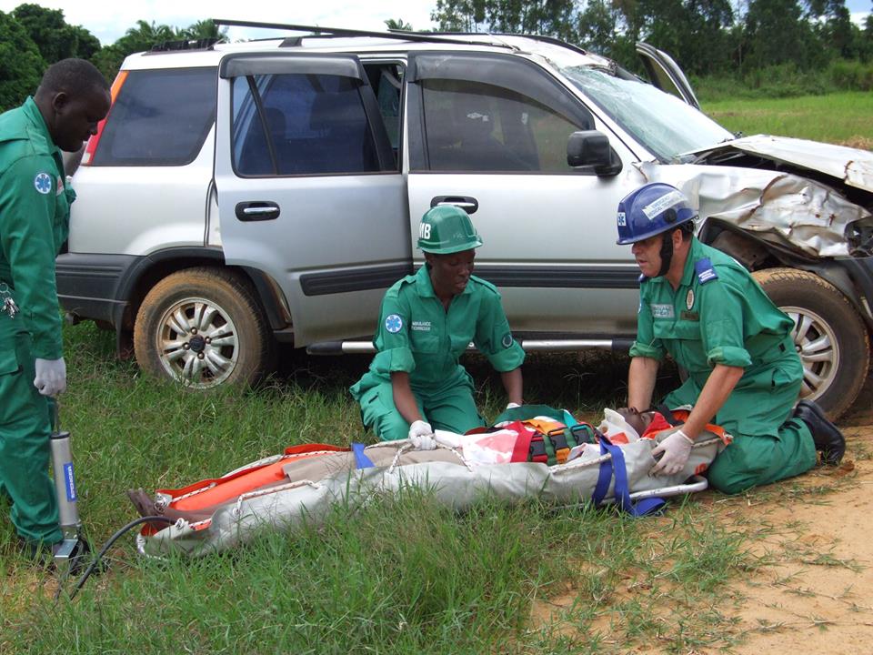
Polytrauma: definition, management, stable and unstable polytrauma patient
With “polytrauma” or “polytraumatized” in medicine we mean by definition an injured patient who presents associated injuries to two or more parts of the body (skull, spine, thorax, abdomen, pelvis, limbs) with current or potential impairment of the functions vital (respiratory and/or circulatory)
Polytrauma, the causes
The cause of multiple traumas is generally linked to a serious car accident but any type of event characterized by a force capable of intervening on multiple points of the same body is capable of resulting in a multiple traumas.
The polytrauma patient is often severe or very severe.
Among the patients who died of polytrauma:
- 50% of polytraumas die within seconds or minutes of the event, due to rupture of the heart or great vessels, laceration of the brainstem or severe cerebral hemorrhage;
- 30% of polytraumas die during the golden hour, due to hemopneumothorax, hemorrhagic shock, liver and spleen rupture, hypoxemia, extradural hematoma, body displacement with worsening of the initial situation or erroneous medical interventions;
- 20% of polytrauma die in the following days or weeks due to sepsis, respiratory problems, cardiac arrest, or acute multiorgan failure (MOF).
The correct, timely and effective intervention of the specific aid allows to increase the chances of survival of the injured person, reducing the risk of secondary damage.
Management of polytrauma
In order to standardize the sequences followed by the team carrying out the rescue, the latter is divided into various phases, called “rings”, which are as follows:
- Preparatory and warning phase – In this phase, the teams are responsible for the correct preparation of the means and facilities that make up the necessary equipment. The operations center is responsible, on the basis of the information in its possession, for alerting the team most suited to the needs.
- Scenario assessment and triage – Upon arrival, each responder is responsible for safety management and risk assessment. The obligations established by law include the identification of a manager and the adoption of personal protective equipment which must be worn correctly and in perfect working order.
- Primary and secondary checks – The necessary assessments of vital functions always correspond to the actions envisaged by the first aid and resuscitation protocols and the alerting of the advanced rescue units (ALS). These controls are mnemonically identified with the acronym ABCDE.
- Communication with the Operations Center – During this phase, in addition to selecting and assigning the destination, the opportunity to call in alternative means of transport or plan a rendezvous with an ALS team is verified.
- Transport with monitoring – During this phase, in addition to the continuous monitoring of the patient’s vital functions, the hospital unit can be provided with information on vital parameters and all those that allow the structure to be prepared to welcome and treat a seriously injured person.
- Healthcare treatment in hospital.
THE RADIO FOR RESCUERS IN THE WORLD? VISIT THE EMS RADIO BOOTH AT THE EMERGENCY EXPO
There is an important and simple rule of thumb for remembering how to provide care for a polytrauma patient, based on the first few letters of the alphabet:
- Airways: or “respiratory tract”, as controlling its patency (i.e. the possibility of air passing through it) represents the first and most contingent condition for the survival of the patient;
- Breathing: or “breath”, intended as “quality of breath”; correlated with the previous point, it is enriched with neurological clinical significance, as some brain lesions give characteristic respiratory patterns (i.e. how much/how/how the patient performs respiratory acts), such as for example Cheyne-Stokes respiration;
- Circulation: or “circulation”, as obviously the correct functioning of the cardiovascular system (and with the two previous points cardio-pulmonary) is essential for survival;
- Disability: or “disability”, particularly important if there is a suspicion of spinal lesion or more generally of the central nervous system, as it may happen that lesions in this district induce a condition of shock which, in its early stages, could not be detectable except by an expert eye, and could “silently” bring the polytraumatized to death (it is no coincidence that sometimes we speak of spinal shock);
- Exposure: or “exposure” of the patient, undressing him in search of any injuries, while safeguarding privacy and temperature (it can also be interpreted as E-nviroment).
First aid, how to deal with a polytrauma
Once in the emergency room, the polytraumatized patient will undergo all the checks that the guidelines for trauma require.
Typically, secondary evaluations for trauma, blood gases, and blood chemistry and blood grouping are done followed by radiological investigations, which will depend on the degree of hemodynamic stability.
Stable polytrauma patient
If a patient is haemodynamically stable, in addition to the basic ecoFAST investigations, x-rays of the chest and pelvis, total body CT investigations can also be performed, both without and with contrast medium, which can highlight neurological lesions and great vessels.
The radiological diagnostic investigations carried out in a severe hemodynamically stable polytrauma are generally:
- FAST ultrasound;
- Chest X-ray;
- pelvis x-ray;
- skull CT;
- cervical spine CT;
- chest CT;
- Abdominal CT.
More in-depth investigations such as angiographies and magnetic resonance may possibly be performed; in particular, MRI is performed on the spine if myelic lesions (of the spinal cord) are suspected, since CT shows the purely bony part of the spine and is not a useful investigation for studying the spinal cord.
MRI can also be performed for the study of the posterior cranial fossa, and in particular for subtle hematomas, which are not satisfactorily highlighted on CT.
X-rays of the limbs are usually performed at the end of the above tests.
The x-ray of the cervical spine is not useful for the in-depth study of bone lesions, as it does not clearly highlight the C1 and C2 vertebrae and would not be sufficient to understand the location of the vertebral fracture.
Unstable polytrauma patient
If a polytraumatized patient is haemodynamically unstable, for example due to active external or internal (or both) bleeding, which has not resolved after the administration of crystalloids, colloids and/or fresh frozen plasma and blood, the patient will not undergo CT investigations, but basic investigations and will subsequently undergo surgery to resolve the complications causing instability.
If a patient arrives in the ED unstable but is subsequently stabilized through therapeutic aids, the trauma team could consider whether to perform more in-depth investigations (such as CT). In particular, the radiological investigations performed in an unstable polytrauma patient (who remains unstable after therapy) generally consist of: -ultrasound (possibly not FAST) -chest X-ray -pelvis X-ray -cervical spine X-ray Cervical spine X-ray is not always performed .
After the investigation
At the end of all the diagnostic investigations, the need for surgery is assessed in the stable patient or possible operations are scheduled for the following days.
The unstable patient is generally taken to the operating room at the end of the basic investigations and will be subjected to more in-depth investigations at the end of the surgery and possibly to secondary surgical operations in the following days.
Polytrauma patients are typically admitted to intensive care units, known simply as “resuscitation” or neurosurgical intensive care units.
Read Also
Emergency Live Even More…Live: Download The New Free App Of Your Newspaper For IOS And Android
Traumatic Injury Emergencies: What Protocol For Trauma Treatment?
Chest Trauma: Symptoms, Diagnosis And Management Of The Patient With Severe Chest Injury
Head Trauma And Brain Injuries In Childhood: A General Overview
Traumatic Pneumothorax: Symptoms, Diagnosis And Treatment
Diagnosis Of Tension Pneumothorax In The Field: Suction Or Blowing?
Pneumothorax And Pneumomediastinum: Rescuing The Patient With Pulmonary Barotrauma
ABC, ABCD And ABCDE Rule In Emergency Medicine: What The Rescuer Must Do
Sudden Cardiac Death: Causes, Premonitory Symptoms And Treatment
Disaster Psychology: Meaning, Areas, Applications, Training
Emergency Room Red Area: What Is It, What Is It For, When Is It Needed?
Emergency Room, Emergency And Acceptance Department, Red Room: Let’s Clarify
Medicine Of Major Emergencies And Disasters: Strategies, Logistics, Tools, Triage
Code Black In The Emergency Room: What Does It Mean In Different Countries Of The World?
Emergency Medicine: Objectives, Exams, Techniques, Important Concepts
Chest Trauma: Symptoms, Diagnosis And Management Of The Patient With Severe Chest Injury
Dog Bite, Basic First Aid Tips For The Victim
Choking, What To Do In First Aid: Some Guidance To The Citizen
Cuts And Wounds: When To Call An Ambulance Or Go To The Emergency Room?
Notions Of First Aid: What A Defibrillator Is And How It Works
How Is Triage Carried Out In The Emergency Department? The START And CESIRA Methods
What Should Be In A Paediatric First Aid Kit
Does The Recovery Position In First Aid Actually Work?
What To Expect In The Emergency Room (ER)
Basket Stretchers. Increasingly Important, Increasingly Indispensable
Nigeria, Which Are The Most Used Stretchers And Why
Self-Loading Stretcher Cinco Mas: When Spencer Decides To Improve Perfection
Ambulance In Asia: What Are The Most Commonly Used Stretchers In Pakistan?
Stretcher: What Are The Most Used Types In Bangladesh?
Travel And Rescue, USA: Urgent Care Vs. Emergency Room, What Is The Difference?


