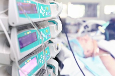
Pressure-controlled Ventilation: using PCV early in a patient’s clinical course may improve outcomes
Positive-pressure ventilation (as opposed to negative-pressure ventilation) has been the basic approach to mechanical ventilation since the late 1950s
The earliest positive-pressure ventilators required the operator to set a specific pressure; the machine delivered flow until that pressure was reached.
At that point, the ventilator cycled into expiration, making the delivered tidal volume dependent upon how quickly the preset pressure was reached.
Anything that caused regional changes in compliance (such as patient position) or resistance (such as bronchospasm) resulted in an undesirable-and often unrecognized-decrease in delivered tidal volumes (and, subsequently, hypoventilation) due to the machine’s premature cycling into the expiratory phase.
Volume-cycled (VC) ventilation was introduced in the late 1960s
This type of ventilation guarantees a consistent, prescribed tidal volume, and has been the method of choice since the 1970s.
Although the tidal volume is uniform with volume-cycled ventilation, changes in compliance or resistance result in an increase in the pressure generated within the lungs.
This can cause barotrauma and volutrauma. In a sense, the solution to the problem of hypoventilation created the problem of excessive pressure/volume.
VENTILATION AND PRESSURE CONTROL
Most newer-generation ventilators are available with the pressure-controlled ventilation (PCV) mode.
In PCV, pressure is the controlled parameter and time is the signal that ends inspiration, with the delivered tidal volume determined by these parameters.
The highest flow is provided at the beginning of inspiration, charging the upper airways early in the inspiratory cycle and allowing more time for pressures to equilibrate.
Flow decelerates exponentially as a function of the rising pressure, and the preset inspiratory pressure is maintained for the duration of the operator-set inspiratory time.
CLINICAL ADVANTAGES
Ventilation/perfusion mismatching often occurs in lungs that have low compliance, as found in adult respiratory distress syndrome (ARDS).
When some lung units have lower compliance than others, gas delivered at a constant flow rate (such as that commonly administered using conventional volume ventilation) follows the path of least resistance.
This results in an uneven distribution of ventilation
When compliance decreases in other lung units, further maldistribution of the breath occurs.
The most compliant lung units become overventilated and the least compliant lung units remain underventilated, causing ventilation/perfusion mismatching.
This often results in high local ventilating pressures and increases the potential for barotrauma.
It has been postulated1 that the high initial peak flow and decelerating inspiratory flow pattern used in PCV can result in recruitment of additional lung units and improved ventilation of alveoli (with prolonged time constants).
This decelerating flow waveform results in more laminar airflow at the end of inspiration, with a more even distribution of ventilation in lungs with markedly different resistance values from one region of the lung to another.2
Waveform analysis allows the clinician to optimize inspiratory time, further reducing ventilation/perfusion mismatching.
The ideal inspiratory time allows both inspiratory and expiratory flows to reach 0 L/min during mechanical breaths.
If the inspiratory time for mechanical breaths is too short, the ventilator cycles into the expiratory phase before inspiratory pressures have adequate time to equilibrate.
This results in a reduced inspired tidal volume.
By lengthening the inspiratory time in very small increments, it is possible to increase the delivered tidal volume and to increase alveolar ventilation.
Caution must be exercised, however, to avoid increasing inspiratory time too much; if it is too long, the expiratory flow does not reach 0 L/min (baseline) before the ventilator cycles into the inspiratory phase.
This indicates (but does not quantify) the presence of intrinsic positive end-expiratory pressure (PEEP), or autoPEEP.
If the inspiratory time is lengthened to the point at which autoPEEP is created, a reduced tidal volume can result.
One method used to reach the optimal inspiratory time is to increase the inspiratory time in 0.1-second intervals until the exhaled tidal volume decreases.
At this point, the inspiratory time should be decreased 0.1 second and maintained.3
Another possible hazard of setting an inspiratory time that is too long is hemodynamic compromise due to increased intrathoracic pressure.
PCV usually results in a higher mean airway pressure.
Some investigators have associated this increase in intrathoracic pressure with hemodynamic compromise, as characterized by decreased cardiac output4 and a significantly reduced cardiac index.5
On occasion (particularly with a high preset respiratory rate), zero flow cannot be reached on inspiration or expiration, creating a paradox.
The clinician must decide whether to increase inspiratory or expiratory time to achieve the most desirable tidal volume and hemodynamic results for the particular patient.
The shapes of ventilator waveforms can exhibit significant changes as the condition of the diseased lung changes, sometimes in a very short time.
For this reason, careful and consistent monitoring of the flow-time curve is important.
Monitoring tidal volume is also important.
No tidal volume guarantee is present in PCV compared to volume ventilation.
Patients may be hypo- or hyperventilated as changes in compliance and resistance occur.
ADVANTAGES OF PCV (pressure-controlled ventilation)
Improved V/Q Match
PCV has been most commonly used in patients, such as those with ARDS, who have significantly reduced lung compliance characterized by high ventilating pressures and worsening hypoxemia despite a high fraction of inspired oxygen (Fio2) and level of PEEP.1,3,4,6-9
By delivering the mechanical breath with an exponentially decelerating flow pattern, PCV allows pressures to equilibrate across the lung units during a preset time, resulting in significantly reduced pressures and in improved distribution of ventilation.
This lowers the risk of barotrauma attributable to the high pressures often required to ventilate these patients.
Studies1,6-9 suggest PCV improves arterial oxygenation and oxygen delivery to the tissues.
One possible explanation for this improved oxygenation is that PCV causes an increase in alveolar recruitment, with reductions in shunting and dead space ventilation.3
Because improved oxygenation has been associated with increased mean airway pressure,2,6,9 this mean pressure level should be recorded prior to the conversion to PCV; adjustments should be made in PEEP levels and inspiratory time (if possible) to maintain a consistent mean airway pressure.
Some authors also suggest that autoPEEP is closely related to oxygenation5 and recommend using autoPEEP as a primary control variable for oxygenation.10
Extremely high airway resistance, as found in severe bronchospasm, results in serious ventilation/perfusion mismatching.
The high airway resistance causes very turbulent gas flow, generating high peak pressures and very poor distribution of ventilation.
The exponentially decelerating waveform of PCV creates more laminar airflow at the end of inspiration.
Administering the breath over a fixed period of time “splints” the airways open so a more even distribution of ventilation to the lung units that participate in gas exchange can occur.
Improved Synchrony
Occasionally a patient’s inspiratory flow demand exceeds the flow-delivery capability of the ventilator in VC ventilation. When the ventilator is set to deliver a fixed flow pattern, as in conventional volume ventilation, it does not adjust inspiratory flow to accommodate the flow needs of the patient. In PCV, the ventilator matches flow delivery and patient demand, making mechanical breaths much more comfortable and often decreasing the need for sedatives and paralytics.
Lower Peak Airway Pressures
The same tidal volume setting, delivered by PCV versus VC, will result in a lower peak airway pressure.
This is a function of the shape of the flow waveform and may explain the lower incidence of barotrauma and volutrauma with PCV.
INITIAL SETTINGS
For PCV, the initial inspiratory pressure can be set as the volume-ventilation plateau pressure minus PEEP.
Respiratory rate, Fio2, and PEEP settings should be the same as those for volume ventilation. Inspiratory time and inspiratory to expiratory (I:E) ratio are determined based on the flow-time curve.
When PCV is used for high inspiratory flow and high airway resistance, however, the inspiratory pressure should be started at a relatively low level (usually < 20 cm H2O) and inspiratory time should be relatively short (usually < 1.25 seconds in adults) to avoid excessively high tidal volumes.
In changing any of the ventilator settings, careful consideration must be given to the effect the change will have on other variables.
Changing inspiratory pressure or inspiratory time will change the delivered tidal volume.
Changing the I:E ratio changes the inspiratory time, and vice versa.
When changing respiratory rate, keep the inspiratory time constant so as not to change the tidal volume, although this will alter the I:E ratio.
Always observe the flow-time curve when making changes (for immediate determination of the effect of the change on breath delivery dynamics).
Watch for oxygenation changes when manipulating any variables that might change the mean airway pressure.
Increasing PEEP while maintaining a constant peak airway pressure-that is, decreasing the inspiratory pressure the same amount as the increase in PEEP-will cause a decrease in delivered tidal volume.
Conversely, a decrease in PEEP with a constant peak airway pressure will result in an increase in delivered tidal volume.
TRANSITION TO PCV (pressure-controlled ventilation )
At our institution, an early transition to PCV for individuals at risk for pulmonary complications (ARDS, aspiration pneumonia, and the like) appears to have improved outcomes by preventing some of the hazards associated with mechanical ventilation, such as barotrauma.
Future studies should examine the role of PCV early in a patient’s clinical course, when respiratory failure may be less severe and the overall physiologic state may be better.
Improvement following the initiation of PCV is not always immediate.
Although reduced peak airway pressure is frequently observed immediately, other improvements may appear only after several minutes or hours.
For example, there is often an initial decrease in oxygen saturation because previously underventilated units begin to participate in gas exchange, causing immediate ventilation/perfusion mismatching.
In the absence of signs of hemodynamic compromise, it is suggested that one leave the patient in PCV until full stabilization has been allowed to occur.
Inverse I:E ratios are not always necessary.
Early published reports6,8,10 indicated that inverse I:E ratios were always to be used with PCV.
More recent published reports3,5 have questioned the utility of this concept.
A great deal has been written about the effects of inverse I:E ratios on hemodynamic parameters such as cardiac output and pulmonary capillary wedge pressure.
Some investigators1,6,8 have found PCV to have little or no effect on hemodynamic variables, while others4,5 suggest significant effects on these parameters.
One recent study3 found the use of an inverse I:E ratio is not universally necessary.
Any adverse hemodynamic effects of inverse I:E ratios will vary from patient to patient.
Whether or not inverse ratios are used, individual hemodynamic parameters should be monitored to the extent possible, and corrective action should be taken if any adverse effects occur.
For example, high autoPEEP will require an increase in E time with either a reduction in respiratory rate or an increase in the I:E ratio (from 1:1 to 1:1.5).
CONCLUSION
Current microprocessor ventilators have given us the ability to revisit an old form of ventilation with much greater safety and efficiency.
Studies on PCV are becoming increasingly common in the medical literature, and favorable results are being reported across the full spectrum of patients, from pediatric through adult populations.
To keep up with the PCV information explosion, and apply this ventilatory mode safely and efficiently, RCPs should have a thorough understanding of the fundamental concepts of PCV.
REFERENCES:
- Abraham E, Yoshihara G. Cardiorespiratory effects of pressure controlled ventilation in severe respiratory failure. Chest. 1990;98:1445-1449.
- Marik PE, Krikorian J. Pressure-controlled ventilation in ARDS: a practical approach. Chest. 1997;112:1102-1106.
- Howard WR. Pressure-control ventilation with a Puritan-Bennett 7200a ventilator: application of an algorithm and results in 14 patients. Respiratory Care. 1993;38:32-40.
- Chan K, Abraham E. Effects of inverse ratio ventilation on cardiorespiratory parameters in severe respiratory failure. Chest. 1992;102:1556-1661.
- Mercat A, Graini L, Teboul JL, Lenique F, Richard C. Cardiorespiratory effects of pressure-controlled ventilation with and without inverse ratio in the adult respiratory distress syndrome. Chest. 1993;104:871-875.
- Lain DC, DiBenedetto R, Morris SL, Nguyen AV, Saulters R, Causey D. Pressure control inverse ratio ventilation as a method to reduce peak inspiratory pressure and provide adequate ventilation and oxygenation. Chest. 1989;95:1081-1088.
- Sharma S, Mullins RJ, Trunkey DD. Ventilatory management of pulmonary contusion patients. Am J Surg. 1996;172:529-532.
- Tharrat RS, Allen RP, Albertson TE. Pressure controlled inverse ratio ventilation in severe adult respiratory failure. Chest. 1988;94:7855-7862.
- Armstrong BW, MacIntyre NR. Pressure-controlled inverse ratio ventilation that avoids air trapping in the adult respiratory distress syndrome. Crit Care Med. 1995;23:279-285.
- East TD, Bohm SH, Wallace CJ, et al. A successful computerized protocol for clinical management of pressure control inverse ratio ventilation in ARDS patients. Chest. 1992;101:697-710.
READ ALSO:
Emergency Live Even More…Live: Download The New Free App Of Your Newspaper For IOS And Android
Endotracheal Intubation: What Is VAP, Ventilator-Associated Pneumonia
The Purpose Of Suctioning Patients During Sedation
Supplemental Oxygen: Cylinders And Ventilation Supports In The USA
Basic Airway Assessment: An Overview
Respiratory Distress: What Are The Signs Of Respiratory Distress In Newborns?
EDU: Directional Tip Suction Catheter
Suction Unit For Emergency Care, The Solution In A Nutshell: Spencer JET
Airway Management After A Road Accident: An Overview
Tracheal Intubation: When, How And Why To Create An Artificial Airway For The Patient
What Is Transient Tachypnoea Of The Newborn, Or Neonatal Wet Lung Syndrome?
Traumatic Pneumothorax: Symptoms, Diagnosis And Treatment
Diagnosis Of Tension Pneumothorax In The Field: Suction Or Blowing?
Pneumothorax And Pneumomediastinum: Rescuing The Patient With Pulmonary Barotrauma
ABC, ABCD And ABCDE Rule In Emergency Medicine: What The Rescuer Must Do
Multiple Rib Fracture, Flail Chest (Rib Volet) And Pneumothorax: An Overview
Internal Haemorrhage: Definition, Causes, Symptoms, Diagnosis, Severity, Treatment
Assessment Of Ventilation, Respiration, And Oxygenation (Breathing)
Oxygen-Ozone Therapy: For Which Pathologies Is It Indicated?
Difference Between Mechanical Ventilation And Oxygen Therapy
Hyperbaric Oxygen In The Wound Healing Process
Venous Thrombosis: From Symptoms To New Drugs
What Is Intravenous Cannulation (IV)? The 15 Steps Of The Procedure
Nasal Cannula For Oxygen Therapy: What It Is, How It Is Made, When To Use It
Nasal Probe For Oxygen Therapy: What It Is, How It Is Made, When To Use It
Oxygen Reducer: Principle Of Operation, Application
How To Choose Medical Suction Device?
Ambulance: What Is An Emergency Aspirator And When Should It Be Used?
Ventilation And Secretions: 4 Signs A Patient On A Mechanical Ventilator Requires Suctioning


