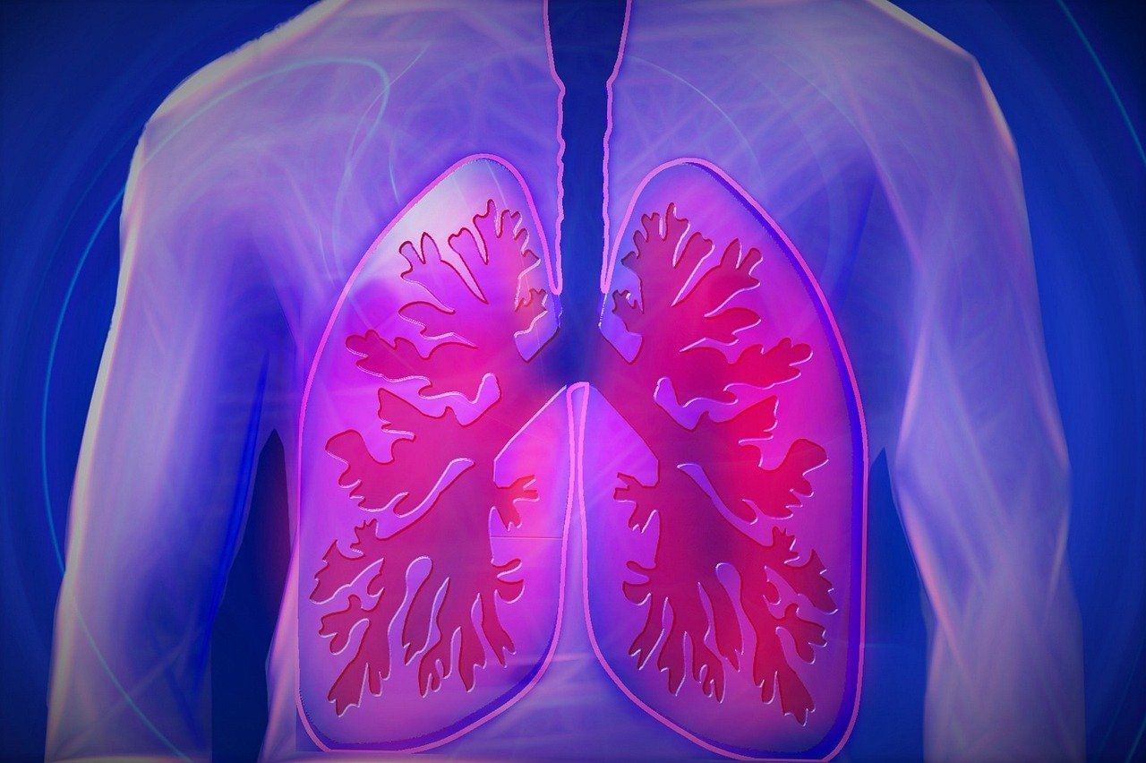
Pulmonary emphysema: what it is and how to treat it. The role of smoking and the importance of quitting
Pulmonary emphysema is one of the diseases caused by cigarette smoking (but not only), which leads to breathing difficulties
Figures presented during World No Tobacco Day, the annual occasion dedicated to raising awareness of the importance of quitting smoking, show that in 2022 almost 1 in 4 Italians (24.2% of the population) will be a smoker: a percentage up by 2 percentage points compared to the pre-pandemic since 2006.
Smoking, as is now well known, is an important (if not the main) risk factor for the development of many diseases (such as cancers).
These include pulmonary emphysema
It is estimated to affect about 210 million people worldwide and may cause the death of 3 million individuals each year.
In the past, pulmonary emphysema was more common among men, who were heavy smokers.
In recent years, however, the scenario has changed: even women smokers, now more numerous than in the past, are affected by pulmonary emphysema and at the same time, much more often than men, also by chronic obstructive bronchopathy, a disease related to emphysema, as we shall see below.
Early intervention, especially to prevent the decline in lung function, is not only possible, but necessary.
What is pulmonary emphysema and the different types
Emphysema is a disease of the pulmonary alveoli: the tissue from which they are composed deteriorates with a reduction in their ability to exchange oxygen and carbon dioxide with the blood.
The alveolar tissue is destroyed, severely reducing the useful surface area for gas exchange: once destroyed, the 7 alveoli can no longer return to their former state, they are irreparably damaged.
From a morphological point of view, several types of pulmonary emphysema are classified:
- centrolobular (or centroacinar) pulmonary emphysema, the most common form in smokers;
- panlobular (or panacinous) pulmonary emphysema;
- paraseptal pulmonary emphysema;
- irregular pulmonary emphysema.
What are the causes
There can be many causes, but, in the West, smoking (tobacco consumption) is the main cause (90% of cases).
Causes therefore include:
- cigarette smoking, including passive smoking
- inhalation of toxic substances;
- being the child of smoking mothers during pregnancy;
- air pollution;
- recurrent respiratory infections;
- prematurity and low birth weight;
- Alpha 1-antitrypsin deficiency.
- Cigarette smoke and respiratory inflammation
Inhaling toxic vapours, such as those found in cigarette smoke, damages cells and promotes an inflammatory state.
This results in the elimination of damaged cells and, at the same time, the inhibition of natural repair mechanisms, leading to the development of emphysema.
The lungs lose elasticity, the alveoli rupture, creating large air spaces that reduce the surface area necessary for the body to exchange oxygen and carbon dioxide.
This process, associated with the chronic inhalation of harmful substances, such as cigarette smoke, often occurs together with a state of chronic inflammation of the airways, called chronic bronchitis, leading to a complex pathology known as chronic obstructive bronchopathy.
Let us not forget that continuous infections of the lower airways also create inflammation and, by increasing mucus secretion, can contribute to the course of the disease.
Pulmonary emphysema – the symptoms
One of the earliest symptoms of pulmonary emphysema is certainly shortness of breath (or dyspnoea), which gets progressively worse: at first it appears when doing intense physical exertion, then when performing everyday tasks such as climbing stairs, and finally even at rest.
In addition, the progressive destruction of the alveoli and pulmonary capillaries, as well as the lack of oxygen, can lead to an increase in pulmonary arterial pressure, which can lead to right heart failure (this is referred to as ‘pulmonary heart disease’).
Finally, patients with emphysema have a higher probability of experiencing pneumothorax, i.e. the formation of a breach in the lung tissue that leads to a collapse of the lung.
In addition to dyspnoea and heart failure, they may experience:
- a dry cough with chronic expectoration;
- fatigue;
- heart problems;
- fever;
- cyanosis of the lips and nails.
How the diagnosis is made: tests to be done
Emphysema usually affects smokers around the age of 50 and presents itself insidiously with shortness of breath during physical exertion, which is often attributed by the patient to age or sedentariness.
Unfortunately, patients often only visit their doctor after an episode of bronchitis after which they are unable to breathe as before, by which time the disease is already quite advanced.
For this reason, it is very important for General Practitioners to be proactive in looking for the disease in their smoking patients over 40 years of age by investigating whether they have a frequent cough or have noticed shortness of breath during physical activities.
Constant coughing and shortness of breath: the first signs to look out for
It is therefore very important for a patient who smokes to consult their doctor if they have
- a cough almost every day for at least 3 months of the year for 2 consecutive years
- shortness of breath for physical activities that did not bother him a year earlier.
The family doctor will be able to collect a correct anamnesis and objective examination and then organise the appropriate examinations, possibly with the help of a pulmonary specialist to establish the best therapy and prevention of complications.
Spirometry
The most important test for diagnosing chronic obstructive pulmonary disease is spirometry, which will show a picture of obstruction to expiratory flow.
It is a simple, non-invasive, inexpensive examination that is easy to perform and interpret.
The subject simply has to blow hard into an instrument that measures the flow of air starting with a deep inhalation.
Normally, a healthy person should be able to empty between 70-80% of all the air they can expel in the first second of the manoeuvre.
Patients with an airway obstruction or loss of lung elasticity, as occurs in emphysema, take much longer.
This obstruction typically responds little or not at all to the administration of a bronchodilator drug.
Further functional tests
Once the picture has been identified, confirmation of emphysema could be made by performing other functional tests, such as global spirometry and alveolar-capillary diffusion, which assess both lung hyperinflation and loss of efficiency of gas exchange typical of emphysema.
Computed tomography of the lung can also show areas of alveolar destruction at a very early stage.
For more severe cases, pulse oximetry measurement will give information on blood oxygenation and, if necessary, arterial haemogasanalysis, the taking of blood from the wrist), will be useful to check the correct gas exchange within the alveoli, the level of oxygen in the blood and predict proper lung function.
How to treat pulmonary emphysema
There is no specific treatment that can restore lost respiratory function, the only thing that can change the natural history of emphysema is to stop smoking.
Quitting smoking modifies the accelerated decline in lung function, slowing the progressive course of the disease.
Unfortunately, quitting the smoking habit is not easy, but today we have smoke-free centres that can both help against nicotine addiction and provide psychological support to counteract psychological dependence.
This combined approach has significantly improved the success in smoking cessation in motivated people.
In addition to smoking cessation, patients should be encouraged to adopt a healthy lifestyle, maintain regular physical activity and protect themselves against infection with flu and pneumococcal vaccination.
Drug therapy for pulmonary emphysema
Other therapies available are bronchodilators, which are used to reduce expiratory flow limitation by reducing lung hyperinflation and improving shortness of breath.
Anti-inflammatory drugs are also used which, in some patients, can reduce bronchial obstruction and prevent bronchial flare-ups and thus preserve pulmonary function.
These drugs can alleviate symptoms and thus also improve patients’ quality of life.
Antibiotics, on the other hand, are only indicated during flare-ups of chronic bronchitis or for pneumococcal pneumonia.
Other therapies
For patients with severe forms leading to respiratory insufficiency, supplementary oxygen for at least 18 hours a day is indicated to help prevent ‘pulmonary heart disease’ (right heart failure).
On the other hand, for all patients whose breathlessness interferes with their daily life, respiratory rehabilitation is indicated.
The latter consists of a multidisciplinary programme aimed at improving exercise tolerance with physiotherapeutic interventions to strengthen limb and respiratory muscles, as well as providing educational and nutritional support to help patients manage their chronic disability.
Possible complications
The most frequent complications are flare-ups, defined as episodes of worsening shortness of breath and coughing that are sometimes severe enough to endanger the patient’s life.
These episodes can further impair lung function, leading to a higher stage of severity.
The causes of flare-ups are often viral, sometimes bacterial infections or pneumonia.
Sometimes, they can also complicate with heart attacks or episodes of heart failure.
A greater effort is therefore needed to seek out patients with this disease at the earliest possible stage, starting secondary prevention of smoking cessation immediately, initiating appropriate drug therapies and interventions aimed at modifying patients’ lifestyles, so that the evolution of the disease can be countered from its onset.
Read Also:
Emergency Live Even More…Live: Download The New Free App Of Your Newspaper For IOS And Android
Oxygen-Ozone Therapy: For Which Pathologies Is It Indicated?
Hyperbaric Oxygen In The Wound Healing Process
Venous Thrombosis: From Symptoms To New Drugs
What Is Intravenous Cannulation (IV)? The 15 Steps Of The Procedure
Nasal Cannula For Oxygen Therapy: What It Is, How It Is Made, When To Use It


