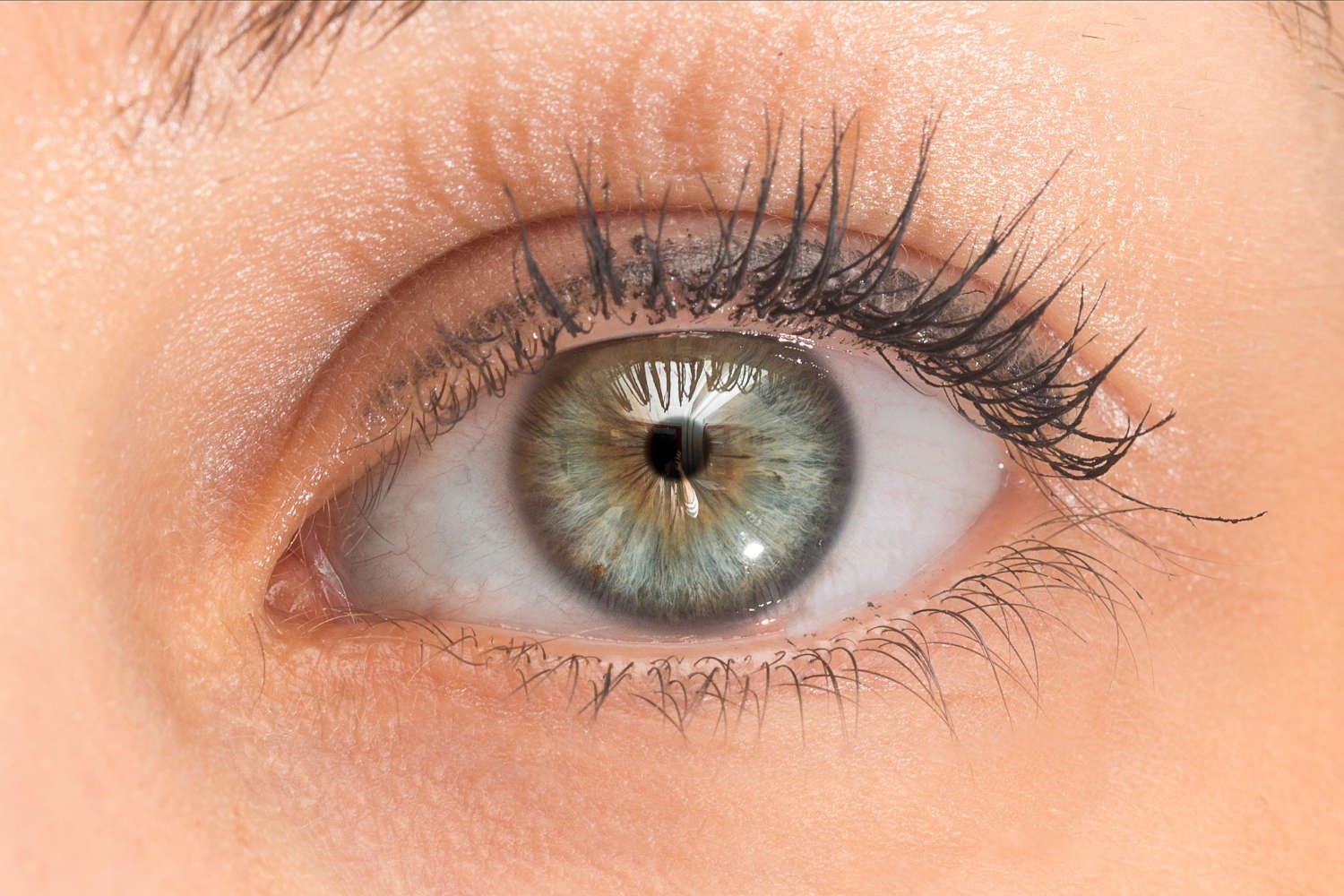
Pupillary reflex to light: mechanism and clinical significance
Pupillary reflex to light (also called photomotor reflex) is a nerve reflex that modulates the diameter of the pupil in response to the intensity of light reaching the retina
This is due to two opposing mechanisms
- increase in light stimulus -> narrowing of the pupil (miosis) allowing less light to enter the retina;
- decrease in ambient light -> dilation of the pupil (mydriasis) which allows more direct light to enter the retina.
The healthy eye is able to switch quickly from miosis to mydriasis when passing from a very bright environment to a dark one and vice versa, think for example when driving a car in broad daylight and entering or exiting a dark tunnel.
Put simply, such a system allows us not to be dazzled in the presence of excessive ambient light and at the same time to ‘capture’ the little light available in dark environments, allowing us the best possible quality of night vision.
Physiological mechanism of the pupillary reflex
- The optic nerve constitutes the afferent pathway of the pupillary reflex: it perceives incoming light.
- The oculomotor nerve constitutes the efferent pathway: it controls the pupil constrictor muscles.
In detail, the pupillary reflex pathway involves four neurons in succession:
- The retinal ganglion cells, which transport information from the photoreceptors to the optic nerve. This reaches the pretectal nucleus in the superior midbrain.
- From here, a second neuron reaches the Edinger-Westphal nucleus.
- From the Edinger-Westphal nucleus a third neuron forms the ipsi- and contralateral oculomotor nerves, which reach the ciliary ganglia.
- Finally, the fourth neuron forms the short ciliary nerve, which innervates the pupil constrictor muscle.
Clinical significance of the pupillary reflex
In addition to regulating the amount of light entering the eye, the pupillary reflex to light provides a useful diagnostic tool.
It allows a physician or ophthalmologist to assess the integrity of the sensory and motor functions of the eye.
Under normal conditions, the pupils of both eyes respond identically to a light stimulus, regardless of which eye is stimulated.
Light entering one eye produces a constriction of both the pupil of the same eye (direct response) and that of the unstimulated eye (consensual response). The comparison of these two responses in both eyes is useful for localising a lesion.
For example:
- a direct response in the right pupil without a consensual response in the left one indicates a possible problem in the motor connection to the left pupil (as a result of damage to the oculomotor nerve or the brainstem Edinger-Westphalnel nucleus);
- the lack of response to light stimulation of the right eye if both eyes respond normally when the left is stimulated indicates damage to the sensory afferent pathway from the right eye (the retina or the right optic nerve).
Normally, both pupils should constrict when light is directed into only one of the eyes.
The absence or abnormality of the pupillary reflex may be due – in addition to damage to the optic nerve or oculomotor nerve – to brainstem death or drugs that depress the central nervous system, such as barbiturates.
Read Also:
Emergency Live Even More…Live: Download The New Free App Of Your Newspaper For IOS And Android
First Aid For Dehydration: Knowing How To Respond To A Situation Not Necessarily Related To The Heat
Hydration: Also Essential For The Eyes
What Is Aberrometry? Discovering The Aberrations Of The Eye
Red Eyes: What Can Be The Causes Of Conjunctival Hyperemia?
Ectopia Lentis: When The Lens Of The Eye Shifts
Chalazion: What It Is And How To Treat This Inflammation Of The Eyelid
Patient Conditions: How To Assess Reflexes
Contusions And Lacerations Of The Eye And Eyelids: Diagnosis And Treatment


