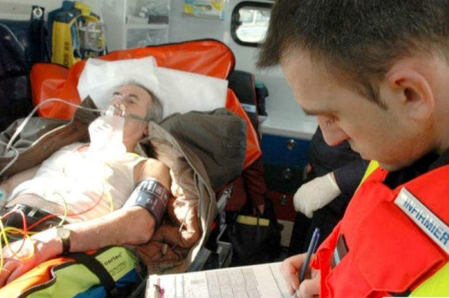
Respiratory Assessment: A Brief Overview
Respiratory Assessment: one of the most critical scenarios you as a paramedic will respond to is respiratory emergencies
Respiratory distress can be linked to many underlying medical problems, so it must be recognized immediately, treated proficiently, and monitored continuously.
But like any medical emergency, treatment begins with a good assessment.
So, let’s review the fundamentals of a proper respiratory assessment.
About Respiratory Assessment: Identifying Respiratory Distress
A patient’s complaint of difficulty breathing is subjective.
As with pain, you must rely on what your patient tells you to gauge his or her status.
But there are also visible signs of respiratory distress that you as a provider must recognize.
They include:
A) General appearance
B) Vital signs
- Respiratory rate
- Pulse
- Oxygen saturation
C) Capnography
D) Level of consciousness
E) Obvious signs of distress
F) Positioning
G) Anxiety
H) Tachycardia
I) Tachypnea
- Skin color and moisture
L) Diaphoretic?
M) Pale or cyanotic?
- Signs of imminent respiratory arrest
N) Decreasing level of consciousness
O) Patient tiring/exhaustion
P) Cyanosis—a late sign and unreliable
Respiratory Assessment, How Hard Are They Working?
Assessing work of breathing is another vital step in identifying respiratory distress.
If your patient is having to work to breath, something is wrong.
Here are five key signs that your patient is working too hard:
- The use of accessory muscles and obvious retractions
- Inability to speak smoothly
- Inability to lie flat
- Diaphoresis
- Agitation and restlessness that will decline to loss of consciousness
Identify the Pattern
Pattern of breathing can also be a good indicator of an underlying problem, so be sure to observe and document the patient’s respiratory pattern, especially if it changes.
- Kussmaul’s respirations: fast and deep labored breathing, often punctuated by sighs, associated with metabolic acidosis
- Cheyne–Stokes: a cyclical pattern of breathing of progressive increased rate and depth of respirations followed by periods of apnea; often associated with overdose, acidosis, and increased ICP
- Apneustic breathing: prolonged periods of gasping inspiration followed by brief, ineffective expiration
- Hyperventilation: increased rate and depth of respirations associated with anxiety, fever, exertion, acid–base imbalance, or damage to the midbrain.
- Bradypnea: abnormally slow rate of respiration associated with drug or alcohol ingestion, central nervous system lesions (both traumatic and non-traumatic), metabolic disorders, and fatigued patients
- Apnea: the absence of respirations
- Agonal respirations: an abnormal pattern that can be slow, shallow, deep, or gasping
Listen to Your Patient
Lung sounds are critical to a proper respiratory assessment.
They can indicate what type of underlying disorder is affecting your patient.
They include:
- Wheezes: indicating a constricted airway
- Crackles: also called rales; heard when collapsed airways or alveoli pop open or when there is mucus in the airway
- Rhonchi: associated with mucus in the airway
- Stridor: a “seal bark” often accompanying infection, swelling, trauma, disease, or a foreign body
Treatment begins with assessment, and no emergency is more critical than those involving the respiratory tract.
Early identification is key, so stay alert for the signs and symptoms of respiratory distress and always conduct a thorough assessment.
Read Also:
Emergency Live Even More…Live: Download The New Free App Of Your Newspaper For IOS And Android
Pressure-Controlled Ventilation: Using PCV Early In A Patient’s Clinical Course May Improve Outcomes
Endotracheal Intubation: What Is VAP, Ventilator-Associated Pneumonia
The Purpose Of Suctioning Patients During Sedation
Supplemental Oxygen: Cylinders And Ventilation Supports In The USA
Basic Airway Assessment: An Overview
Respiratory Distress: What Are The Signs Of Respiratory Distress In Newborns?
EDU: Directional Tip Suction Catheter
Suction Unit For Emergency Care, The Solution In A Nutshell: Spencer JET
Airway Management After A Road Accident: An Overview
Tracheal Intubation: When, How And Why To Create An Artificial Airway For The Patient
What Is Transient Tachypnoea Of The Newborn, Or Neonatal Wet Lung Syndrome?
Traumatic Pneumothorax: Symptoms, Diagnosis And Treatment
Diagnosis Of Tension Pneumothorax In The Field: Suction Or Blowing?
Pneumothorax And Pneumomediastinum: Rescuing The Patient With Pulmonary Barotrauma
ABC, ABCD And ABCDE Rule In Emergency Medicine: What The Rescuer Must Do
Multiple Rib Fracture, Flail Chest (Rib Volet) And Pneumothorax: An Overview
Internal Haemorrhage: Definition, Causes, Symptoms, Diagnosis, Severity, Treatment
Assessment Of Ventilation, Respiration, And Oxygenation (Breathing)
Oxygen-Ozone Therapy: For Which Pathologies Is It Indicated?
Difference Between Mechanical Ventilation And Oxygen Therapy
Hyperbaric Oxygen In The Wound Healing Process
Venous Thrombosis: From Symptoms To New Drugs
What Is Intravenous Cannulation (IV)? The 15 Steps Of The Procedure
Nasal Cannula For Oxygen Therapy: What It Is, How It Is Made, When To Use It
Nasal Probe For Oxygen Therapy: What It Is, How It Is Made, When To Use It
Oxygen Reducer: Principle Of Operation, Application
How To Choose Medical Suction Device?
Ambulance: What Is An Emergency Aspirator And When Should It Be Used?
Ventilation And Secretions: 4 Signs A Patient On A Mechanical Ventilator Requires Suctioning


