Semeiotics of heart failure: the Valsalva Manoeuvre (tachycardia and vagus nerve)
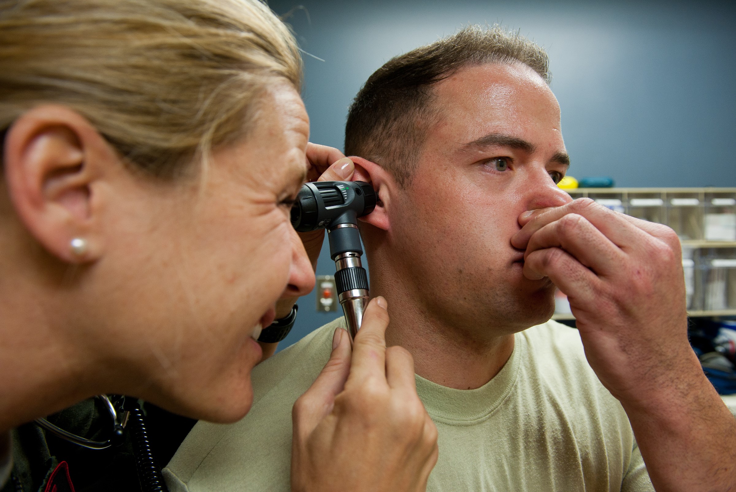
The Valsalva manoeuvre (MV), named after the physician Antonio Maria Valsalva, is a forced compensation manoeuvre of the middle ear, mainly used in medicine, especially in the field of cardiology, but also in the field of diving
What does the Valsalva manoeuvre consist of?
The Valsalva manoeuvre consists of a relatively deep inhalation followed by a forced exhalation with the glottis closed lasting approximately 10 seconds.
What is the glottis?
The ‘glottis’ is the upper segment of the larynx at the vocal cords, located below the epiglottis and above the cricoid cartilage.
The glottis is, in simple terms, the opening of the larynx and corresponds to the natural space that can form between the vocal cords and their respective arytenoid cartilages; it is not a permanent and fixed space since it is affected by the activities and movements of the larynx: during breathing, the glottis takes the form of a triangle, while during phonation (voice emission), the glottis becomes a thin line interposed between the vocal cords.
The glottis has three functions: it enables correct phonation; it isolates the respiratory system from the digestive system, allowing food to pass into the oesophagus and air into the trachea.
What is the Valsalva manoeuvre used for?
Initially, this manoeuvre was used to remove suppuration and foreign bodies from the ear.
Subsequently, the focus shifted to the haemodynamic changes produced by its execution, which proved useful in the diagnostic process of numerous pathological conditions, cardiological and otherwise.
It is also useful in controlling tachycardia.
Valsalva manoeuvre in tachycardia
The Valsalva manoeuvre is taught by cardiologists to patients suffering a paroxysmal tachycardia crisis in order to stop it, as the vagus nerve (X cranial nerve) is stimulated, thus causing a parasympathetic vagal stimulation that slows down the heart rate.
The dynamics of MV involves four phases:
- tension onset phase,
- tension phase,
- release phase,
- recovery phase.
Normally, phase I is characterised, during exhalation with the glottis closed, by the increase in intrathoracic pressure and systolic arterial pressure due to compression of the aorta.
Subsequently, during phase II, there is a decrease in venous return and systolic arterial pressure secondary to the persistence, at intrathoracic level, of a positive pressure.
Simultaneously, there is an increase in heart rate.
During the subsequent phases of relaxation and recovery, the rapid reduction in intrathoracic pressure triggers a series of physiological compensation mechanisms.
Specifically, the rapid change in the blood volume in the pulmonary vascular system leads to an abrupt reduction in systolic blood pressure (phase III) and, subsequently, the increase in cardiac output, peripheral vasoconstriction due to sympathetic hyperactivity and the reduction in heart rate lead to an increase in systolic blood pressure (phase IV).
Is the Valsalva Manoeuvre still useful?
MV has been widely used in ‘classical’ semeiotics for the evaluation of patients with heart failure and for a more thorough assessment of cardiac murmurs.
The advent of more modern imaging methods such as echocardiography has reduced the use of this manoeuvre in clinical practice.
However, it still represents a valuable aid in the echocardiography laboratory in the assessment of left ventricular diastolic function, in the evaluation of the extent of left ventricular outflow obstruction in hypertrophic cardiomyopathy, and in the diagnosis of patency of the foramen ovale (PFO) for the evaluation of the associated right-left shunt.
Furthermore, MV retains a discrete utility in the classical semeiotic evaluation of numerous clinical cardiovascular conditions such as the diagnosis of systolic heart murmurs, autonomic dysfunction, arrhythmias and heart failure.
Read Also
Emergency Live Even More…Live: Download The New Free App Of Your Newspaper For IOS And Android
Performing The Cardiovascular Objective Examination: The Guide
Heart Diseases And Alarm Bells: Angina Pectoris
Fakes That Are Close To Our Hearts: Heart Disease And False Myths
Sleep Apnoea And Cardiovascular Disease: Correlation Between Sleep And Heart
Myocardiopathy: What Is It And How To Treat It?
Venous Thrombosis: From Symptoms To New Drugs
Cyanogenic Congenital Heart Disease: Transposition Of The Great Arteries
Heart Rate: What Is Bradycardia?
Consequences Of Chest Trauma: Focus On Cardiac Contusion
Heart Murmur: What Is It And What Are The Symptoms?
Branch Block: The Causes And Consequences To Take Into Account
Cardiopulmonary Resuscitation Manoeuvres: Management Of The LUCAS Chest Compressor
Supraventricular Tachycardia: Definition, Diagnosis, Treatment, And Prognosis
Identifying Tachycardias: What It Is, What It Causes And How To Intervene On A Tachycardia
Myocardial Infarction: Causes, Symptoms, Diagnosis And Treatment
Aortic Insufficiency: Causes, Symptoms, Diagnosis And Treatment Of Aortic Regurgitation
Congenital Heart Disease: What Is Aortic Bicuspidia?
Atrial Fibrillation: Definition, Causes, Symptoms, Diagnosis And Treatment
Ventricular Fibrillation Is One Of The Most Serious Cardiac Arrhythmias: Let’s Find Out About It
Atrial Flutter: Definition, Causes, Symptoms, Diagnosis And Treatment
What Is Echocolordoppler Of The Supra-Aortic Trunks (Carotids)?
What Is The Loop Recorder? Discovering Home Telemetry
Electrocardiogram: Initial Procedures, ECG Electrode Placement And Some Tips
What Is The Electrocardiogram (ECG)?
ECG: Waveform Analysis In The Electrocardiogram
What Is An ECG And When To Do An Electrocardiogram
ST-Elevation Myocardial Infarction: What Is A STEMI?
ECG First Principles From Handwritten Tutorial Video
ECG Criteria, 3 Simple Rules From Ken Grauer – ECG Recognize VT
The Patient’s ECG: How To Read An Electrocardiogram In A Simple Way
ECG: What P, T, U Waves, The QRS Complex And The ST Segment Indicate
Electrocardiogram (ECG): What It Is For, When It Is Needed
Stress Electrocardiogram (ECG): An Overview Of The Test
What Is The Dynamic Electrocardiogram ECG According To Holter?
Full Dynamic Electrocardiogram According To Holter: What Is It?
Cardiac Rhythm Restoration Procedures: Electrical Cardioversion
Cardiac Holter, The Characteristics Of The 24-Hour Electrocardiogram
Peripheral Arteriopathy: Symptoms And Diagnosis
Endocavitary Electrophysiological Study: What Does This Examination Consist Of?
Cardiac Catheterisation, What Is This Examination?
Echo Doppler: What It Is And What It Is For
Transesophageal Echocardiogram: What Does It Consist Of?
Paediatric Echocardiogram: Definition And Use


