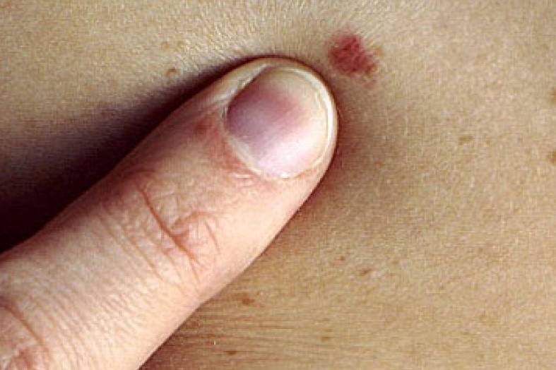
Skin cancers: prevention and care
The skin is a body organ like any other, such as the liver, kidney, lung, heart. However, it has a special characteristic that makes it unique, it is visible
Tumours can affect the skin, like all organs, with the great opportunity of being able to diagnose them early, or prevent them, given precisely by the visibility of the skin organ.
Skin tumours, classification of skin neoformations
Skin tumours are divided into two major groups: epithelial tumours, epitheliomas, and melanocytic tumours, melanoma with its clinical variants (superficial, nodular, acral melanoma and lentigo maligna).
The mortality and aggressiveness of the latter group is far higher than that of epitheliomas.
Melanoma was considered a rare neoplasm until a few years ago, but today it is growing steadily in all countries.
Its incidence has increased more than all other cancers, second only to lung cancer in women (up 30% in the last 10 years).
As indicated by the World Health Organisation – WHO, approximately 132,000 new cases of melanoma are diagnosed worldwide every year.
In Mediterranean countries, the incidence is 3-5 cases per year per 100,000 inhabitants and is slightly higher in the female population than in the male population (7 and 6 per 100,000 per year, respectively).
In our country there are 1500 deaths from melanoma out of 7000 cases diagnosed each year.
Melanoma originates from melanocytes, the skin cells that produce melanin, the main pigment in the skin
It accounts for 4% of skin cancers and is responsible for 80% of cancer deaths in this organ, which occur at the late metastatic stage involving other organs such as the lung, brain and lymph nodes.
Only one in five cases, however, has an advanced form, thanks in part to prevention campaigns and increasingly early diagnosis of the disease thanks to today’s new diagnostic methods.
The skin is a body organ, like the liver, kidney, lung, heart
Individuals at greatest risk are those with a family history, high number of nevi, previous melanoma.
Other risk factors are phototype I – II (blond hair, light-coloured eyes, etc.), chronic exposure to artificial UV radiation (tanning lamps), immunosuppression.
A major study by the International Agency for Research on Cancer (IARC) showed that exposure to tanning lamps, if it occurs under the age of 30, increases the risk of melanoma by 75%.
This resulted in UV radiation being placed in August 2009 in class I of carcinogens, the highest alert, like cigarette smoking.
In addition, several studies on the correlation between intense sun exposure and sunburn during childhood and adolescence have shown a more than double risk of developing cancer in the presence of sunburn at a young age.
It is important to remember that melanoma can arise on healthy skin or on a pre-existing acquired or congenital melanocytic nevus.
Therefore, photoprotection should not only be carried out on nevi but on all exposed skin.
Diagnosis at an early stage (initial melanomatous transformation in situ) guarantees survival equal to the general population.
Therefore education of the general population in annual nevi screening has a positive prognosis.
The skin is made up of layers and in the superficial layers (epidermis) there are no blood or lymph vessels.
In the early stages the melanoma disease, so-called in situ, is located here and has no chance of metastasising.
The aim must be precisely to diagnose it at this stage or, even better, before malignant transformation at the time of dysplasia or atypia that precedes neoplasia.
The general control of nevi should take place annually with a complete evaluation of the entire skin surface, indicating to the patient where melanocytic lesions are, especially in locations that escape daily observation and are perhaps not known (retroauricular region, plantar and interdigital spaces of the feet, back, genitals especially in women, scalp, visible oral and ocular mucosa, etc.).
Of fundamental importance is the so-called mapping of nevi with digital dermoscopy using a suitable instrument
This is a modern non-invasive diagnostic method that makes it possible to map out the body nevi and assess their characteristics, cataloguing those at risk of transformation that will then be kept under control with re-evaluations at a distance (3, 6, 8, 12 months), established on the basis of the degree of dermoscopic atypia found, or possibly removed.
Melanocytic lesions can change over time, so mappings are performed at a distance from the initial one, to see if the lesions change morphology, otherwise a single mapping would be performed in a lifetime.
In addition, as long as we ‘live in our skin’, new lesions may appear every year, but these need to be mapped and checked annually.
The new mapping instruments make it possible to create a photographic archive of nevi to ensure precisely an objective comparison of lesions at a distance and not on the basis of a vague memory of the patient or doctor.
Atypical lesions, or those suspected to be neoplastic, must always be surgically removed in an outpatient procedure and must always be subjected to histopathological analysis for microscopic diagnostic definition.
The patient must be educated to annual mapping and periodic self-examination, every 3-4 months, self-performed by observing the entire skin surface, especially in the sites of rare self-observation, sometimes with the help of a family member or a mirror.
This is intended to anticipate the annual periodic visit if sudden and marked changes in a nevus are noted.
It is recommended to check for any asymmetry of the nevus
Simply dividing the nevus into two parts with a line should present symmetry in terms of colour, edges, size, as well as checking the growth of the nevus.
The patient should not notice the growth of the lesion, in fact an approximately millimetric growth over years is physiological and not noticeable, while a centimetric growth over a short period of time should always be reported to the dermatologist who will dermoscopically assess the lesion.
Finally, a uniform but very dark, black shade (hyperpigmentation) warrants further evaluation of the lesion.
Thus, the patient simply needs to observe asymmetry, rapid growth and hyperpigmentation.
Read Also
Emergency Live Even More…Live: Download The New Free App Of Your Newspaper For IOS And Android
Melanoma: Prevention And Dermatological Examinations Are Essential Against Skin Cancer
Nail Melanoma: Prevention And Early Diagnosis
Dermatological Examination For Checking Moles: When To Do It
What Is A Tumour And How It Forms
Rare Diseases: New Hope For Erdheim-Chester Disease
How To Recognise And Treat Melanoma
Moles: Knowing Them To Recognise Melanoma
Skin Melanoma: Types, Symptoms, Diagnosis And The Latest Treatments
Nevi: What They Are And How To Recognise Melanocytic Moles
Bluish Color Of Baby’s Skin: Could Be Tricuspid Atresia
Skin Diseases: Xeroderma Pigmentosum
Basal Cell Carcinoma, How Can It Be Recognised?
Autoimmune Diseases: Care And Treatment Of Vitiligo
Epidermolysis Bullosa And Skin Cancers: Diagnosis And Treatment
SkinNeutrAll®: Checkmate For Skin-Damaging And Flammable Substances
Healing Wounds And Perfusion Oximeter, New Skin-Like Sensor Can Map Blood-Oxygen Levels
Psoriasis, An Ageless Skin Disease
Psoriasis: It Gets Worse In Winter, But It’s Not Just The Cold That’s To Blame
Childhood Psoriasis: What It Is, What The Symptoms Are And How To Treat It
Topical Treatments For Psoriasis: Recommended Over-The-Counter And Prescription Options
What Are The Different Types Of Psoriasis?
Phototherapy For The Treatment Of Psoriasis: What It Is And When It Is Needed
Skin Diseases: How To Treat Psoriasis?


