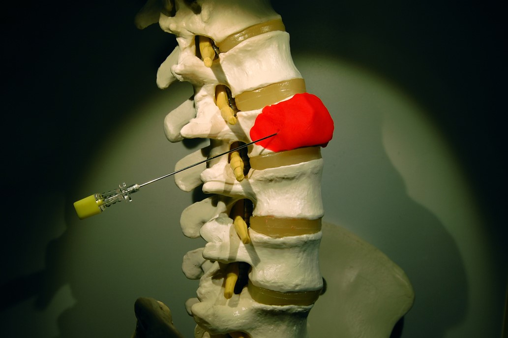
Spinal biopsy: what it is, how it is performed and what risks it presents
Spinal biopsy is a diagnostic test that involves taking a small portion of bone from the spinal column
What is a spinal biopsy?
A biopsy of the vertebral column is based on taking a small portion of bone from the paraspinal soft tissues (anterior and posterior), the soma and posterior vertebral arches, the intervertebral discs, the epidural space and the conjugate foramina.
The procedure is considered effective in the characterisation of vertebral, disc and paraspinal soft tissue lesions.
It has an accuracy rate of around 80-95%, with very high reliability in the study of osteolytic lesions.
What is the purpose of spinal biopsy?
The vertebral biopsy is performed to check for the presence of neoplasms (vertebral or paraspinal), infections and metabolic bone diseases.
How is the vertebral biopsy performed?
The procedure is performed under local anaesthesia and uses fine needles.
Three types of techniques are used to perform the procedure: ‘Tandem’, with the insertion of two needles of different calibre in parallel, one for local anaesthesia and the other for biopsy.
Coaxial’, with the insertion of needles of different calibre one inside the other to reach deep lesions.
Single-needle’ with a mandrel (thin metal wire that is inserted into the needle to prevent it from occluding) inside, used for vertebral and soft tissue biopsies.
During the thirty minutes following the procedure, the patient must be kept lying down and under observation.
After this time has elapsed without any complications appearing, he can be discharged.
Who can perform the test?
The vertebral biopsy is considered a safe method.
However, it must be performed with caution in the presence of coagulation disorders, pregnancy, local infections at the biopsy site (osteomyelitis and spondylodiscitis), inability to access the disc space due to extensive and solid vertebral fusions or neurological complications.
Is spinal biopsy a painful and/or dangerous test?
The vertebral biopsy is performed under local anaesthesia, so the pain, even if slight, is usually well tolerated.
Complications associated with performing this test are quite rare, occurring in 0.2 per cent of cases, and include: paravertebral haematoma, iatrogenic nerve root lesions, pneumothorax, vertebral osteomyelitis, excessive bleeding and spondylodiscitis.
Neurological injuries occur in 0.08% and death in 0.02% of cases.
Read Also
Emergency Live Even More…Live: Download The New Free App Of Your Newspaper For IOS And Android
Fusion Prostate Biopsy: How The Examination Is Performed
Echo- And CT-Guided Biopsy: What It Is And When It Is Needed
What Is Needle Aspiration (Or Needle Biopsy Or Biopsy)?
What Is Echocolordoppler Of The Supra-Aortic Trunks (Carotids)?
What Is The Loop Recorder? Discovering Home Telemetry
Cardiac Holter, The Characteristics Of The 24-Hour Electrocardiogram
Peripheral Arteriopathy: Symptoms And Diagnosis
Endocavitary Electrophysiological Study: What Does This Examination Consist Of?
Cardiac Catheterisation, What Is This Examination?
Echo Doppler: What It Is And What It Is For
Transesophageal Echocardiogram: What Does It Consist Of?
Venous Thrombosis: From Symptoms To New Drugs
Echotomography Of Carotid Axes
What Is A Liver Biopsy And When Is It Performed?
Abdominal Ultrasound: How It Is Performed And What It Is Used For
What Is Retinal Fluorangiography And What Are The Risks?
Echodoppler: What It Is And When To Perform It


