
Symptoms and causes of umbilical hernia pain
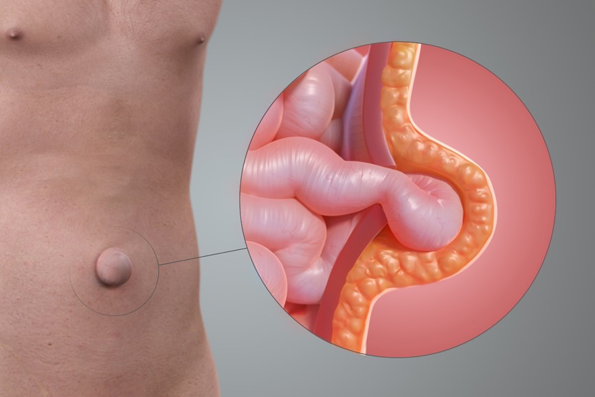
Umbilical hernia is the leakage from the abdominal cavity, through the umbilicus, of part of the intestine, intestinal fat (omentum) and/or the sac that covers the intestine (hernia sac)
In general, it is a pathology that is already evident to the naked eye, however it may be physically less evident, but it generates pain and discomfort.
How umbilical hernia manifests itself
Umbilical hernia generally presents itself without any particular clinical manifestations, apart from the visibility of a swelling in the area of the umbilicus, which becomes more swollen when undergoing physical exertion, while it shrinks and recedes when lying down or with manual manoeuvres.
Umbilical hernia, symptoms to watch out for
However, in more advanced forms of the pathology, there may be certain symptoms to pay attention to:
- pain: this may arise after exertion or, for example, from a small hernial hole from which, however, a considerable portion of the intestine has escaped. If constant and violent, the pain is an alarm bell that must be heeded, and one should go to the emergency room as soon as possible;
- purplish discolouration: the hernia, as it pushes towards the skin of the umbilicus, makes it more delicate and thin, giving it a purplish discolouration that demonstrates the progression of the disease. However, if accompanied by intense and constant pain, this discolouration is another factor that requires immediate medical attention.
Complications of hernia
In the vast majority of patients, umbilical hernia remains an asymptomatic or mildly disabling condition, but in some cases, mainly among adults, it can lead to very serious complications:
- incarcerated hernia: the leaked tract of viscera remains trapped on the outside of the abdominal wall and cannot re-enter, even when lying down or when pushed manually, leading to the serious risk of a strangulated hernia and intestinal obstruction;
- strangulated hernia: the intestine, which is unable to re-enter the abdominal cavity, is strangled on the outside due to the lack of blood flow;
- intestinal occlusion: the contents inside the intestine remain stuck in the section outside the abdominal wall and are unable to move forward, as is the case under normal conditions, to be expelled, with major surgical consequences.
Umbilical hernia in infants
Umbilical hernia is a very frequent pathology in newborns in which it can present itself as a congenital or neonatal hernia: the umbilical cord, severed at the level of the umbilicus, continues to protrude slightly under the skin of this area, leaving a small space of communication with the abdominal cavity, which is therefore not completely closed.
In the newborn this pathology tends to regress and resolve spontaneously.
It should be noted, however, that in both children and adults, umbilical hernia can also occur later as a result of certain circumstances.
Causes of umbilical hernia
As mentioned, the most frequent cause of umbilical hernia for infants is incomplete closure of the umbilical canal, while in childhood or adulthood it is facilitated by factors such as
- obesity, also possibly due to diabetes;
- pregnancy;
- chronic obstructive pulmonary disease, which results in chronic coughing;
- smoking, because it increases the risk of cardio-pulmonary diseases and can cause persistent coughing;
- prolonged constipation;
- significant physical exertion.
These risk factors, which under normal conditions do not lead to a herniated pathology, in umbilical hernia sufferers are associated with a weakness of the tissues due to a metabolic problem with the collagen fibres that makes them more susceptible to increased pressure in the abdominal cavity.
Diastasis of the rectus abdominis muscles
An umbilical hernia is often associated with another phenomenon caused by a weakness in the tissues of the abdominal wall: diastasis of the rectus abdominis, i.e. the distancing and thus the creation of a space between the rectus abdominis muscles, which form 2 parallel blocks, present on the right and left sides of the abdominal wall.
This distancing, which is very frequent, for example, after childbirth, creates the effect of a swollen abdomen that remains so despite exercise, which, if inadequate, can even worsen the clinical picture.
Can an umbilical hernia recur?
A question that is often asked is whether the umbilical hernia can recur and resolve.

The professor’s answer is no: it can re-enter temporarily if lying down or if reinserted manually, but it is basically a hole that, except in the case of newborns, cannot close by itself, but can, at best, remain stable.
The treatment of umbilical hernia
The only treatment for an umbilical hernia is surgery.
If the hernia does not cause any problems, does not grow and does not cause pain, the operation is performed on an aesthetic basis, whereas if there is also discomfort and clinical reasons, it also acquires a functional basis.
The operation for every type of hernia
Each hernia has its own characteristics that provide for a more suitable and effective approach:
- umbilical hernia below 1 cm without diastasis of the rectus abdominis: a minimal operation is generally performed under local anaesthesia, in which the contents leaking out of the abdominal wall are placed inside and the herniated hole closed. No prosthesis is required in this case;
- umbilical hernia above 2 cm without diastasis of the rectus abdominis: performed under local, epidural or spinal anaesthesia (depending on the case), the surgery involves the insertion of a prosthesis, so-called ‘nets’ that repair the hole like a patch;
- umbilical hernia of varying size with diastasis of the rectus abdominis: in this case, although the size of the hernia may be less than a centimetre, a procedure under general anaesthesia is usually performed. The abdominal area, which goes from the end of the sternum to 5/6 cm below the navel, is repaired by inserting a mesh, roughly 15×20 cm, behind the correctly repositioned rectus muscles, without contact with the viscera. In this case, treating only the hernia would expose the patient to a recurrence.
Methods for the treatment of umbilical hernia with diastasis of the rectus
Abdominal wall repair surgery in the presence of hernia and diastasis can be performed with different approaches
- the MILA technique (Minimally Invasive Laparotomy Approach): this can be used for normal-weight, i.e. not obese patients, and involves a 5 cm open incision before proceeding with the operation described above;
- robotic surgery: this technique, which is the most modern, involves the same type of operation with mesh positioning, but with a laparoscopic-robotic approach via 4 small holes;
- suprapubic approach: if the patient has an important cutaneous and subcutaneous ptosis, i.e. laxity generating the ‘sagging tummy’ effect, the operation is performed with a suprapubic incision, a sort of extended caesarean section, which allows a mini-abdominoplasty or abdominoplasty to be performed at the same time, thus not only repairing the herniary hole and repositioning the rectus abdominis, but also removing excess skin in the abdominal area.
The post-operative: when to resume your regular life
In minimal operations under local anaesthesia, the patient can generally return to his normal life as early as the day after the operation.
In the case of operations under general anaesthesia, on the other hand, 1/2 day hospitalisation is required, but overall, within 1 week, subjects resume a regular life with sports activities that can be practised gradually as early as 8/9 days later.
There are, therefore, no particular post-operative precautions to follow, except for the use of compression bands that also help drain excess fluid.
Read Also:
Emergency Live Even More…Live: Download The New Free App Of Your Newspaper For IOS And Android
What It Is And How To Recognise Abdominal Diastasis
Chronic Pain And Psychotherapy: The ACT Model Is Most Effective
Hiatal Hernia: What It Is And How To Diagnose It
Percutaneous Discectomy For Herniated Discs
What Is That Swelling? Everything You Need To Know About Inguinal Hernia