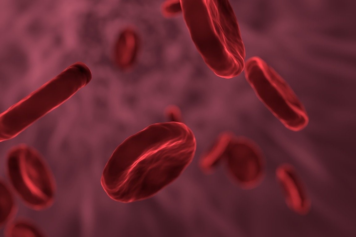
Thalassaemia, an overview
Thalassaemia yes, but it is more correct to speak of thalassaemias, which are hereditary blood diseases involving anaemia, i.e. a decrease in the amount of haemoglobin, a protein contained within red blood cells that has the function of transporting oxygen in the blood
Thalassaemias are a group of haemoglobin defects (haemoglobinopathies) that cause microcytic anaemia, i.e. red blood cells that are smaller than normal (thalassaemia).
Haemoglobin consists of four chains, two alpha chains and two beta chains, each of which contains iron (Fe 2+) and carries oxygen (O2).
Thalassaemias are classified according to the type of altered chain
Two main types can therefore be distinguished:
- Alpha thalassaemia, when the gene that contains the information needed to synthesise the alpha chains is altered and is more common in African individuals or those of African descent;
- Beta thalassaemia or Mediterranean anaemia when the altered gene is the one containing the information needed to synthesise the beta chain and is more frequent in subjects from the Mediterranean area and south-east Asia.
Alpha thalassaemia is caused by alterations in one or more of the four genes that contain the information needed to synthesise the alpha chain of haemoglobin
Most of the genetic alterations that cause alpha thalassaemia are deletions, i.e. losses of a DNA segment of varying length.
However, there are also numerous mutations that can alter the function of the four genes that direct the synthesis of the haemoglobin alpha chains.
There are two genes for the alpha chain (HBA1 and HBA2) on each chromosome number 16, so we all have 4 genes for haemoglobin alpha: two copies of the HBA1 gene and two copies of the HBA2 gene.
Obviously, since we each inherit one chromosome 16 from our mother and the other chromosome 16 from our father, one of the two HBA1 genes is inherited from our mother and the other from our father.
The same applies to the two HBA2 genes.
Subjects with alterations in only one of the 4 genes (Figure 2) are silent carriers of alpha thalassaemia as the other three genes allow the production of almost normal amounts of alpha chains.
Subjects with alterations in 2 of the 4 genes for alpha chain have trait thalassaemia with small red blood cells and moderate anaemia.
Alterations in 3 of the 4 genes lead to a condition of alpha thalassaemia intermedia or haemoglobin H disease manifested by moderate to severe anaemia and thalassaemia.
When all 4 genes for the alpha chains are altered and non-functional, an abnormal haemoglobin is formed (Bart’s haemoglobin) that is unable to transport oxygen to the body, leading to fetal hydrops (excessive accumulation of fluid in the tissues and cavities of the fetus) that is essentially incompatible with life (fetal hydrops with Bart’s haemoglobin).
Beta thalassaemia is by far the most common form of the disease in Italy and along the Mediterranean Sea coastline
It is caused by alterations (mutations) in the gene that contains the information needed to synthesise the beta chain of haemoglobin.
There is one copy of the gene for the beta chain on each of the two chromosomes number 11, so everyone has two copies of the gene.
When only one copy of the gene is altered, the child has trait thalassaemia or thalassaemia minor.
If, on the other hand, the child has inherited two copies of the altered gene from two parents who both have thalassaemic trait, the child has thalassaemia major.
When both parents have the thalassaemic trait, they have a 25% chance of having a child with thalassaemia major with each pregnancy.
If, on the other hand, only one of the parents has the thalassaemic trait, he or she will have a 50% risk of passing on the altered gene to their child each pregnancy, who will then also have the thalassaemic trait.
In the context of alpha thalassaemia:
- The silent carrier condition gives no symptoms and does not even show up on the blood count as there is no anaemia and no thalassaemia either;
- Thalassaemic trait does not usually give symptoms, but can be suspected when a blood count reveals moderate to mild microcytic anaemia;
- Alpha thalassaemia intermedia manifests itself with the symptoms of moderate-to-severe microcytic anaemia with moderate jaundice, liver and spleen enlargement and moderate skeletal changes with skull bone deformities and zygomatic bone protrusion. Statural growth retardation is also common;
- Almost all foetuses with fetal hydrops with Bart’s haemoglobin die in utero or in the first few hours of life unless they are treated immediately in intensive care and transfused.
They present with very severe anaemia, marked enlargement of liver and spleen volume, delayed brain development, skeletal, cardiovascular and urogenital abnormalities.
As for beta thalassaemia:
The condition trait thalassaemia or thalassaemia minor does not usually cause symptoms.
It is usually suspected when a blood count shows mild anaemia and small red blood cells (thalassaemia minor, revealed by a low Mean Corpuscular Volume – MCV).
Thalassaemia major, on the other hand, usually manifests itself within the first or second year of life with symptoms related to severe anaemia and the haemolytic component: feeling of weakness (asthenia), pallor of the skin, jaundice, gallstones, enlarged spleen and liver.
More rarely, skeletal changes occur due to bone marrow hyperplasia, resulting in deformities of the skull bones and protrusion of the zygomatic bone.
Statural growth is also often slowed.
The diagnostic pathway varies depending on the suspected thalassaemic condition:
- Diagnosis as a silent carrier of alpha thalassaemia is usually made in the parents of a child with a more severe form of alpha thalassaemia: blood counts are also usually within normal limits. Diagnosis must be based on the use of molecular tests demonstrating a deletion or mutation in one of the four genes that direct the synthesis of the alpha chains of haemoglobin;
- Thalassaemia trait can be suspected from the results of a haemochromocytometric examination performed for a variety of indications (as a rule, these subjects do not present symptoms) showing moderate anaemia with thalassaemia; the diagnosis must be confirmed by molecular tests;
- In alpha thalassaemia intermedia the diagnostic suspicion comes from the symptoms and the demonstration of moderate to severe microcythaemia; an initial confirmation may come from the demonstration of haemoglobin H in the blood. Molecular tests can highlight deletions or mutations that cause the disease;
- In infants with hydrops fetalis with Bart’s haemoglobin, the diagnosis may come from electrophoretic demonstration or other more sophisticated chromatographic techniques of Bart’s haemoglobin. In this case too, molecular investigations allow the diagnosis to be confirmed.
In alpha-thalassaemias, Hb F and Hb A2 are usually normal, and the diagnosis of thalassaemia by defect in one or two genes can be made by genetic testing.
The diagnostic pathway is obviously different when beta thalassaemia is suspected.
The diagnosis of trait thalassaemia in beta thalassaemia, usually suspected on the basis of the results of a haemochromocytometric examination performed for other reasons, is confirmed by haemoglobin electrophoresis showing high levels of haemoglobin A2.
The diagnosis of thalassaemia major is confirmed by laboratory investigations:
- Haemochromocytometric examination demonstrating severe anaemia of the microcytic type; haemoglobin drops progressively to very low values;
- Peripheral venous blood smear, which is often diagnostic for the presence of many small and pale red blood cells, oddly shaped red blood cells or target red blood cells;
- Increased bilurin, iron and ferritin levels;
- Haemoglobin electrophoresis, where in beta-thalassaemia minor there is an increase in Hb A2, whereas in beta-thalassaemia major, Hb F is increased and also HbA2 over 3%;
- Recombinant DNA methods are now performed to identify the specific molecular defect (genotype), both for prenatal diagnosis and as part of genetic counselling.
Treatment depends on the type and severity.
Silent carriers and individuals with alpha thalassaemia trait and beta thalassaemia minor require no treatment.
Patients with alpha thalassaemia intermedia, fetal hydrops survivors with Bart’s haemoglobin, and children with thalassaemia major require regular and periodic red blood cell transfusions in order to maintain adequate haemoglobin levels according to the individual’s developmental stages and health status.
Since transfused blood contains high amounts of iron, serum ferritin levels must be kept under control: after a certain number of transfusions and if serum ferritin exceeds safe levels, in order to prevent or reduce iron accumulation in the various organs (heart, liver, endocrine glands, etc.), therapy to remove excess iron must be initiated (ferrochelating therapy with drugs such as Deferoxamine, Deferasirox and Deferiprone).
Another therapeutic perspective, although not resolving, is the use of a drug, Luspatercept, the progenitor of the class of erythroid maturation agents.
In some individuals with transfusion-dependent anaemia, including thalassaemic patients, its action of increasing red blood cell production has been observed to reduce transfusion frequency by increasing intervals.
This could lead in selected, responsive cases to fewer transfusions and thus less martial load and accumulation.
Resolutive treatments leading to cure are:
- Haematopoietic stem cell transplantation from an HLA-compatible unaffected sibling, from one of the parents (haploidentical transplantation) or from a bone marrow bank donor;
- LentiGlobin-based beta-thalassaemia gene therapy, which represents the new therapeutic frontier. LentiGlobin is a virus (lentivirus) manipulated in the laboratory in such a way that it transports the haemoglobin beta chain gene into red blood cell precursors taken from the patient. Once the gene has been introduced into the test tube, the precursors, which are now able to synthesise normal haemoglobin beta chains, are transfused back to the patient and enable him or her to produce normal red blood cells.
Read Also
Emergency Live Even More…Live: Download The New Free App Of Your Newspaper For IOS And Android
Mediterranean Anaemia: Diagnosis With A Blood Test
Iron Deficiency Anaemia: What Foods Are Recommended
What Is Albumin And Why Is The Test Performed To Quantify Blood Albumin Values?
What Is Cholesterol And Why Is It Tested To Quantify The Level Of (Total) Cholesterol In The Blood?
Gestational Diabetes, What It Is And How To Deal With It
What Is Amylase And Why Is The Test Performed To Measure The Amount Of Amylase In The Blood?
Adverse Drug Reactions: What They Are And How To Manage Adverse Effects
Albumin Replacement In Patients With Severe Sepsis Or Septic Shock
Provocation Tests In Medicine: What Are They, What Are They For, How Do They Take Place?
What Are Cold Agglutinins And Why Is The Test Performed To Quantify Their Values In The Blood?
Thalassaemia Or Mediterranean Anaemia: What Is It?


