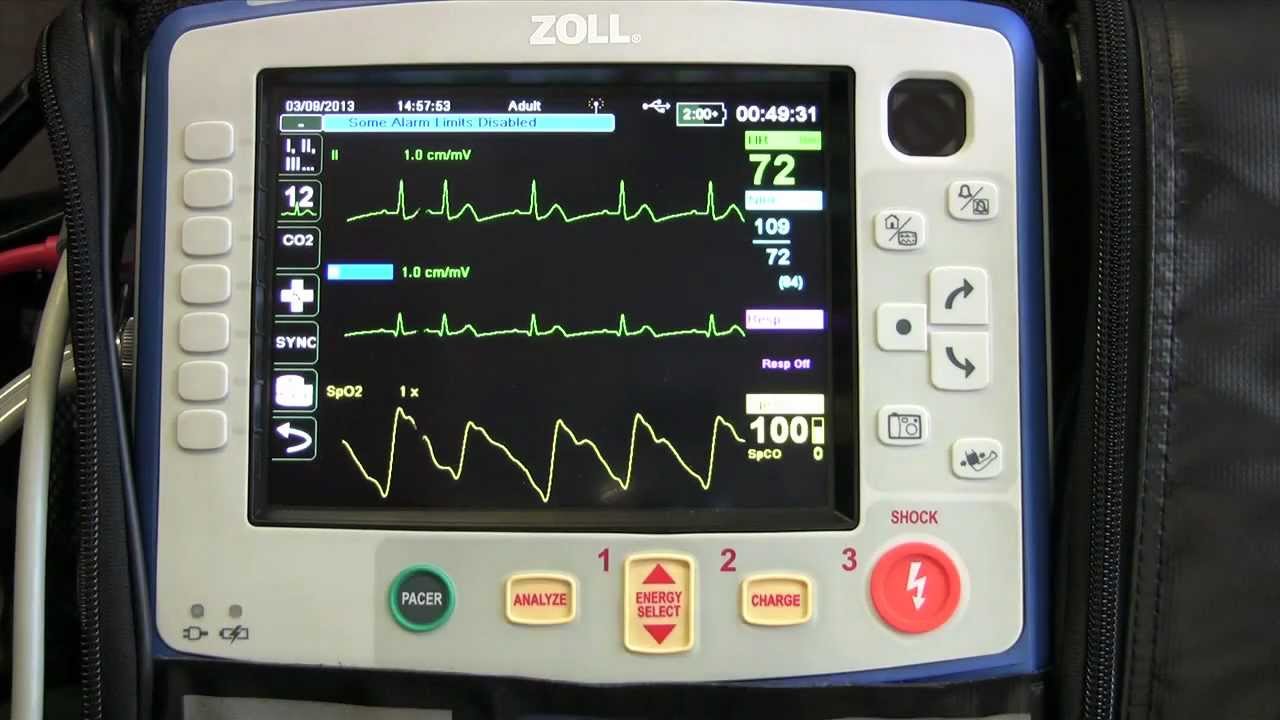
The patient's ECG: how to read an electrocardiogram in a simple way
The electrocardiogram (ECG) tracing is characterised by several traits called positive and negative waves, which repeat at each cardiac cycle and indicate the specific activity of the heart related to the propagation of the cardiac electrical impulse
The normal ECG tracing has a characteristic appearance that changes only in the presence of problems: a given pathology tends to result in a specific alteration at one or more points of the tracing, returning waves that are altered in height, shape or inverted. In this article you will find indications for a basic interpretation of normal and altered electrocardiographic tracing.
For ECG interpretation to be reliable, the electrodes must be positioned correctly: an error in positioning can lead to false-positive results, i.e. result in altered waves indicating pathologies that are not really present.
Accurate reading of an ECG tracing requires a lot of knowledge and experience.
Normal electrocardiogram (ECG) waves, complexes, intervals, tracts and segments
These are defined as:
- positive waves: the waves that are above the isoelectric line;
- negative waves: the waves that are above the isoelectric line.
P wave
This is the first wave generated in the cycle and corresponds to the depolarisation of the atria.
It is small, as the contraction of the atria is not as powerful.
Its duration varies between 60 and 120 ms, and its amplitude (or height) is 2.5 mm or less.
QRS complex
Corresponds to the depolarisation of the ventricles and is formed by a set of three waves that follow one another:
- Q wave: is negative and small, and corresponds to depolarisation of the interventricular septum;
- R wave: is a very high positive peak, and corresponds to depolarisation of the apex of the left ventricle;
- S wave: this is also a small negative wave, and corresponds to depolarisation of the basal and posterior regions of the left ventricle. The duration of the entire complex is between 60 and 90 ms. Atrial repolarisation also occurs in this interval, but is not visible as it is masked by ventricular depolarisation.
T wave
Repolarisation of the ventricles.
It is not always identifiable because it can also be very small in value.
U wave
This is a wave that cannot always be appreciated in a trace, it represents the repolarisation of the Purkinje fibres.
ST Tract (or segment)
This is the distance between the S wave and the start of the T wave, it represents the interval between ventricular depolarisation and the start of ventricular repolarisation (restoration of basic electrical conditions).
Compared to isoelectric, it should be neither above nor below by more than 1 mm in all leads except V1 and V2, in which, however, it should remain below 2 mm.
QT interval
Represents electrical systole, i.e. the time in which ventricular depolarisation and repolarisation occurs.
Its duration varies as the heart rate varies, generally remaining between 350 and 440ms.
PR interval
This is the distance between the onset of the P wave and the onset of the QRS complex; it represents the interval required for atrial depolarisation to reach the ventricles.
It must be between 120 ms and 200 ms in duration (3 to 5 squares).
Interpreting the Adult ECG
Heart rate (HR) and RR interval
Heart rate is defined as the number of heartbeats per minute (bpm) and is related to ventricular rate.
Having a HR of 70 bpm means that 70 contractions of the ventricles occur in one minute.
Obtaining HR from an electrocardiographic trace is quite simple.
The ECG trace is compiled on graph paper, which runs through the electrocardiograph at a rate of 25 mm per second, so five sides of 5 mm squares represent 1 second.
It is therefore easy to imagine how heart rate can immediately be obtained by estimating how much time passes between one cycle and the next (the time between two R peaks is measured, called the RR interval).
Just as an example, if we have a complex every 4 squares of 5 millimetres, this means that our frequency is around 75 beats per minute.
That is, since each 5 mm square corresponds to 0.2 s and, therefore, 4 squares to 0.8 s, we need only divide 60 s (1 minute) by 0.8 s to obtain the frequency of 75 beats per minute.
Or, more simply, we can divide 300 by the number of 5 mm squares between two adjacent R peaks.
Calculating an irregular heart rate
What has just been said applies when the heart rhythm is normal, but in the case of an irregular rhythm, i.e. if you notice that the peaks of the R wave do not occur at regular intervals and are spaced by a variable number of squares, you must count the number of peaks present in six seconds and multiply the result by 10.
This calculation gives an estimate of the heart rate; for example, if in a six-second trace interval you can see seven R waves, you can estimate that the heart beats at the rate of 70 beats per minute (7 x 10 = 70).
Alternatively, you can count the number of QRS complexes present on a trace that is 10 seconds long; multiply this value by 6 to find the number of beats per minute.
Bradycardias and tachycardias
A normal frequency in adults at rest ranges from 60 to 100 bpm.
Higher frequencies are called tachycardias, lower frequencies bradycardias; both can be either physiological (a physiological tachycardia occurs when we exercise, for example, while a physiological bradycardia is typical of professional athletes) or pathological.
Electrocardiogram, rhythm analysis: regular and sinus?
A first assessment is to establish whether the intervals between the R waves are always the same, or do not differ from each other by more than 2 squares.
In this case we can say that the rhythm is regular.
The second assessment relates to the presence and morphology of the P wave: if this is located before the QRS complex and is positive in DII and negative in aVR, then we can define the rhythm as sinus, i.e. the electrical impulse originates from the sinoatrial node (normal condition).
The presence of a negative P wave in DII, must suggest, firstly, a possible inversion of the peripheral electrodes, secondly, a different origin of the impulse than normal (extrasystole and/or atrial tachycardia -TA-).
Sometimes the P wave is not before the QRS complex, but after it: in this case it is linked to retro-conduction of the impulse, which occurs in many arrhythmias, both supraventricular (TPSV) and ventricular (VT).
The presence of an irregular rhythm associated with the absence of a clear P wave, must suggest the most frequently encountered arrhythmia in daily practice: atrial fibrillation (AF).
This is defined as chaotic electrical activity of the atria, resulting in ineffective contraction of the walls and a consequent high probability of clot formation within them.
Another frequently encountered arrhythmia, characterised by a sometimes even regular rhythm and typical sawtooth-like waves (F-waves) is atrial flutter (FLA).
It is caused by an electrical short circuit (re-entry arrhythmia) affecting the atrium. It differs from AF by a greater regularity of the ventricular cycle.
QRS morphology
Normally it should be positive in DI, the amplitude of the R wave should increase from V1 to V6 while the S wave should decrease, duration should be less than 100-120 ms (2.5-3 squares), the Q wave should have a duration of less than 0.04 sec (1 square) and the amplitude should be less than ¼ of the next R wave (Q waves in DIII and aVR are not considered).
Based on the duration of the complex, wide or narrow QRS tachycardias or bradycardias are defined.
When it is narrow (duration less than 100 ms) it indicates normal ventricular conduction.
If it is longer than 120 ms, it is defined as wide and indicates a slowing of conduction, which may be of a specific portion of the conduction system (as in the case of branch blocks), or a sub-Hissian origin of the heart rhythm (junctional or ventricular).
The presence of a wide QRS tachycardia with variable amplitude and morphology from one complex to another is typical of ventricular fibrillation (VF).
This is the arrhythmia that most frequently causes cardiac arrest in association with VT; it is caused by a disorganised electrical activity of the ventricles, resulting in a cessation of mechanical activity.
If immediately before a wide QRS we find a rapid deflection characterised by a vertical line (spike), we are dealing with pacemaker stimulation.
T-wave morphology
When it has the same polarity as the QRS in the peripheral leads and is positive in the precordial leads (or negative from V1 to V3 in young women), it indicates normal ventricular repolarisation. Otherwise it indicates myocardial ischaemia or suffering, ventricular hypertrophy, heart disease).
PR interval, relationship between P waves and QRS complexes
The PR interval expresses the conduction of the impulse through the atrio-ventricular node, the bundle of His, and the left and right branches.
It must be between 120 ms and 200 ms in duration (3 to 5 squares).
When it is shorter, it may be a normal variant (occurring for example in pregnant women) or identify the presence of an atrio-ventricular accessory pathway (ventricular pre-excitation, WPW).
If it is long, it is indicative of a slowing of conduction to the ventricles (atrioventricular blocks or BAV).
Under normal conditions the P:QRS ratio is 1:1, i.e. each P wave, after a constant PR interval, corresponds to a QRS complex and each QRS complex must be preceded by a P wave.
When, on the other hand, we find a P:QRS ratio of 1:2 or 1:many, and a PR interval that has a progressively increasing duration, we are dealing with Atrio-Ventricular Blocks (AVB):
- 1st degree atrioventricular block: prolonged PR
- 2nd degree type I atrioventricular blocks: progressive lengthening of the PR interval until there is no conduction in the ventricle (blocked P i.e. not followed by the QRS)
- 2nd degree type II atrioventricular blocks: the PR interval is normal but conduction is 1:2, 1:3, 1:4, etc.
- 3rd degree atrioventricular blocks or complete block: atrioventricular dissociation, with no constant relationship between P waves and QRS complexes.
In 3rd degree AVB the number of P waves is generally greater than the number of (narrow) QRS.
In the case of ventricular tachycardias, however, the number of QRS complexes (wide) is generally greater than the number of P waves.
QT interval in the electrocardiogram
Expresses the total time of ventricular depolarisation and repolarisation and varies with heart rate; therefore it is more correctly expressed as QTc, i.e. corrected for heart rate. The normal value ranges from 350 to 440 ms.
It is pathological both when it is shorter (short QT syndrome) and when it is longer (long QT syndrome) and in both cases is associated with an increased likelihood of developing ventricular arrhythmias.
ST Tract
Expresses the termination of ventricular depolarisation; it can be found fused with the T wave from V1 to V3 and, with respect to the isoelectric, must be neither above nor below by more than 1 mm in all leads except V1 and V2, in which, however, it must remain below 2 mm.
When a higher than normal superelevation is present, we speak of myocardial injury, i.e. a picture compatible with acute myocardial infarction (AMI).
The location of the superelevation allows the localisation of the infarct and the coronary artery affected by the obstruction:
- an ST-segment elevation in DII, DIII and aVF (with mirror subleveling in DI and aVL) is indicative of inferior myocardial infarction from right coronary artery occlusion;
- ST-segment elevation in DI, V2-V4 (with specular undersegmentation in DII, DIII and aVF) is indicative of anterior myocardial infarction from anterior interventricular branch occlusion.
Read Also:
Emergency Live Even More…Live: Download The New Free App Of Your Newspaper For IOS And Android
Heart Disease: What Is Cardiomyopathy?
Inflammations Of The Heart: Myocarditis, Infective Endocarditis And Pericarditis
Heart Murmurs: What It Is And When To Be Concerned
Broken Heart Syndrome Is On The Rise: We Know Takotsubo Cardiomyopathy
What Is A Cardioverter? Implantable Defibrillator Overview
‘D’ For Deads, ‘C’ For Cardioversion! – Defibrillation And Fibrillation In Paediatric Patients
Inflammations Of The Heart: What Are The Causes Of Pericarditis?
Do You Have Episodes Of Sudden Tachycardia? You May Suffer From Wolff-Parkinson-White Syndrome (WPW)
Knowing Thrombosis To Intervene On The Blood Clot
Patient Procedures: What Is External Electrical Cardioversion?
Increasing The Workforce Of EMS, Training Laypeople In Using AED
Difference Between Spontaneous, Electrical And Pharmacological Cardioversion
What Is Takotsubo Cardiomyopathy (Broken Heart Syndrome)?


